Capture: grabbing the antibody (Protein A)
📍 Where we are: Stop 13 of 21, and the first real purification step. We have a clear tank of liquid full of our medicine plus thousands of unwanted leftovers — now we reach in and pull the antibody out.
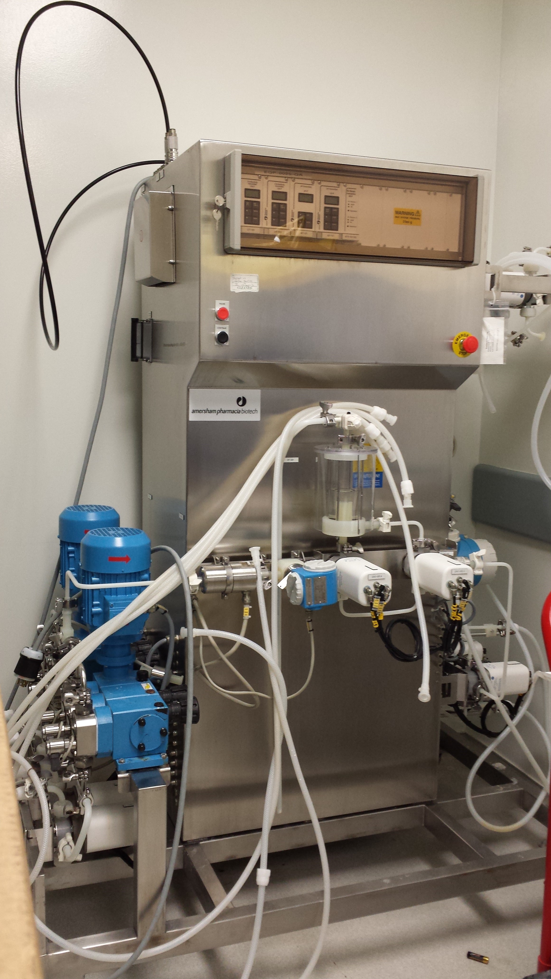 A process chromatography skid packages the pumps, valves, column connections, and detectors that run a capture cycle — the hardware behind the Protein A step in this chapter.
Process chromatography skid. Image by Kitmondo Lab, CC BY 2.0, via Wikimedia Commons.
A process chromatography skid packages the pumps, valves, column connections, and detectors that run a capture cycle — the hardware behind the Protein A step in this chapter.
Process chromatography skid. Image by Kitmondo Lab, CC BY 2.0, via Wikimedia Commons.
After harvest and clarification, we have a clear soup. Our medicine — the antibody (a Y-shaped protein your immune system uses to grab specific targets) — is floating in it, but so are thousands of unwanted leftovers: stray proteins from the factory cells, fragments of DNA, bits of broken cells, leftover nutrients. Capture is the step that fishes out the antibody and throws the great majority of everything else away. It is the single highest-leverage move in the whole purification — one clever step that does most of the work [1].
Imagine a bucket of sand with a few iron nails mixed in. Drag a magnet through it: the nails jump onto the magnet, the sand pours away. Now lift the magnet out and gently switch it off — the nails drop into a clean cup. That magnet is our capture step. The nails are the antibody. The sand is everything we want gone. The only catch: our "magnet" is fussy about exactly which shape it grabs, and we have to nudge it to let go.
What this chapter covers
We will start with the tool — chromatography — and the special "magnet molecule," Protein A, that makes capture so selective. Then we will walk the four-stage cycle (load, wash, elute, clean) and read the chromatogram it produces (a graph of what flows out of the column — defined in full below). We will look at the real beads, real numbers, and real machines used in factories today: how much antibody fits on a litre of resin, what acid releases it, why the resin has to survive being scrubbed with caustic, and how much it costs. We will close with the modern, never-stops version of capture — continuous multi-column chromatography — and how it sets up the very next step, viral inactivation (a dedicated step that kills off any stray viruses, which the acidic conditions of capture conveniently begin to do).
What actually happens
Affinity chromatography and Protein A: the selective grabber
The tool we use is called chromatography — a way to separate a mixture by pumping it through a tube and letting some things stick while others flow past.
Picture a tall, narrow tube called a column. It is packed with millions of tiny porous beads, together called the resin. Liquid is pumped in the top and trickles down through the beads to the bottom. As it passes, the beads can grab certain molecules and hold them, while everything else flows straight through. The beads are porous like sponges, so the liquid does not just slide past their surface — it soaks into a vast internal honeycomb, which is where most of the grabbing happens.
For capture we use a special trick called affinity chromatography — the beads are coated with a grabber molecule named Protein A, and that grabber is picky. Protein A came originally from the surface of the bacterium Staphylococcus aureus, where it helps the microbe dodge the immune system by latching onto antibodies. We borrow that exact talent. It clamps onto the bottom "stem" of the Y — a region called the Fc — that the most common class of human antibodies shares. (Antibodies come in a few families called classes; IgG is the workhorse class nearly all mAb — monoclonal antibody — drugs belong to) [2].
Here is the honest caveat behind the "grabs only antibodies" analogy: Protein A mainly grips IgG by its Fc stem, and it does not bind every antibody class or subclass equally well (human IgG3, for instance, binds poorly). For the IgG-type molecules that make up essentially the entire approved-mAb landscape, though, it is wonderfully selective — almost nothing else in the soup fits the grip. That selectivity is the whole reason capture works so well.
The four-phase cycle: load, wash, elute, clean
This is the cycle in its simplest form. In a real plant it is bracketed by an equilibration step: the column is first rinsed to the binding pH and conductivity (conductivity is just a measure of how much dissolved salt the liquid carries) — typically around pH 7.0 to 7.4 at low salt — so the antibody will stick. (pH is the acidity scale, running 0 to 14: 7 is neutral, lower numbers are more acidic, higher are more alkaline. The antibody grips Protein A best near neutral and lets go when we turn the liquid acidic — the whole cycle below is built on that fact.) And the single "wash" below is usually two or more washes. Here is the simplified cycle, step by step:
- Load. Pump the clarified soup through the column at a deliberately gentle pace. The antibody binds the Protein A beads; the host cell proteins (HCPs — stray proteins from the factory cells), DNA, and cell debris mostly do not stick, so they flow out the bottom to waste. The flow is slow on purpose: the liquid lingers in the column for a residence time of roughly 3 to 6 minutes so the antibody has time to diffuse into the bead pores and find a Protein A grabber before it would otherwise wash past — this is why we drag the magnet through the sand slowly rather than yank it, since the nails need a moment to find it. We do not load to the brim: the column is loaded to a target challenge (grams of antibody per litre of resin) set deliberately below its capacity — often defined at a small breakthrough percentage — to leave a safety margin against losing product out the bottom. Breakthrough means antibody escaping unbound out the bottom because the resin is filling up; "1 or 10 percent breakthrough" is the small fraction we allow to slip past before we stop loading.
- Wash. Push clean buffer (a gentle, pH-controlled salt-water solution) through to rinse away anything loosely hanging on. In practice this is usually two or more washes — an equilibration-buffer rinse plus an intermediate wash (often elevated salt, or a mild additive such as polysorbate or arginine — gentle, antibody-friendly chemicals that help pry loose impurities without disturbing the bound antibody) tuned specifically to dislodge HCP that co-binds to the antibody or the resin backbone (the bead surface the grabber molecules sit on). That intermediate wash is the single biggest real-world lever on HCP clearance, and designing it is why one Protein A step can clear 2 to 3 log of HCP rather than 1 — where each "log" is a tenfold cut, so 2 to 3 log means a hundred- to thousand-fold reduction. The antibody stays gripped.
- Elute. Switch the liquid to a mild acid — a low-pH buffer, often around 50 to 100 mM (millimolar, a measure of concentration) of acetate, citrate, or glycine (all gentle, antibody-safe acids) at roughly pH 3.5, which is mildly acidic, well below the near-neutral binding pH of 7.0 to 7.4. The low pH protonates key residues (adds positive charge to the few amino-acid spots where the antibody and Protein A touch), so those spots now repel rather than cling — weakening the binding interactions so the antibody releases and flows out as a concentrated, far purer stream. The change is reversible — it loosens the grip without destroying the ligand (another name for the Protein A grabber molecule), which is why the column survives to capture again. This release is called elution. Elution can be a single low-pH step or a shallow pH gradient; a gradient can begin to separate monomer from aggregate — single, correctly folded antibody molecules versus clumps of several stuck together, and aggregates are an unwanted impurity that can trigger an immune reaction, so we want as few as possible — and so reduce the aggregate load carried forward into the low-pH hold (the brief acid step that follows, which is the viral-inactivation step described at the end of this chapter).
- Clean and reuse. The column is scrubbed — usually with a sodium hydroxide (caustic) solution — and re-equilibrated so it can capture the next load. This cleaning is called clean-in-place (CIP), and the same caustic doubles as the sanitization step that keeps bioburden (the population of stray microbes) down, since the ligand cannot be autoclaved (sterilized with pressurized steam — the heat would destroy the protein grabber).
One quiet but crucial detail: the antibody does not enjoy sitting in acid. The eluate is the most aggregate-prone moment of the entire train, because the product is concentrated and acidic at the same instant. So the freshly eluted pool is neutralized almost immediately — its pH nudged back up to roughly 5 to 7 — to protect the antibody from clumping or unfolding while it waits for the next step.
A lower pH means more acidic, and the whole cycle is really just three pH moves:
| pH set-point | When | What it does |
|---|---|---|
| ~7.0 to 7.4 (low salt) | Equilibrate & load | Antibody grips Protein A |
| ~3.5 (mild acid) | Elute | Grip loosens; antibody releases |
| ~5 to 7 | Neutralize after elution | Protects the antibody from clumping in acid |
In one sentence: bind by the Fc stem, wash impurities away, release with mild acid, then clean and reuse — and watch the chromatogram to know it worked.
Throughout, a sensor watches the liquid leaving the column and draws a graph called a chromatogram — a line that stays low and flat while impurities pour out unbound, then climbs into a tall, sharp peak exactly when the pure antibody is eluted. That peak is the moment we collect. That UV trace, and the decision of which slice of the peak to keep, is the first rich stream of process data this step throws off — each reading becomes a timestamped, tagged data point in the batch's data shadow (the growing digital twin of the physical lot, the running record every measurement adds to); the companion data book follows exactly how such a signal is born and travels in where data is born, the data shadow, and the lifecycle of a data point.
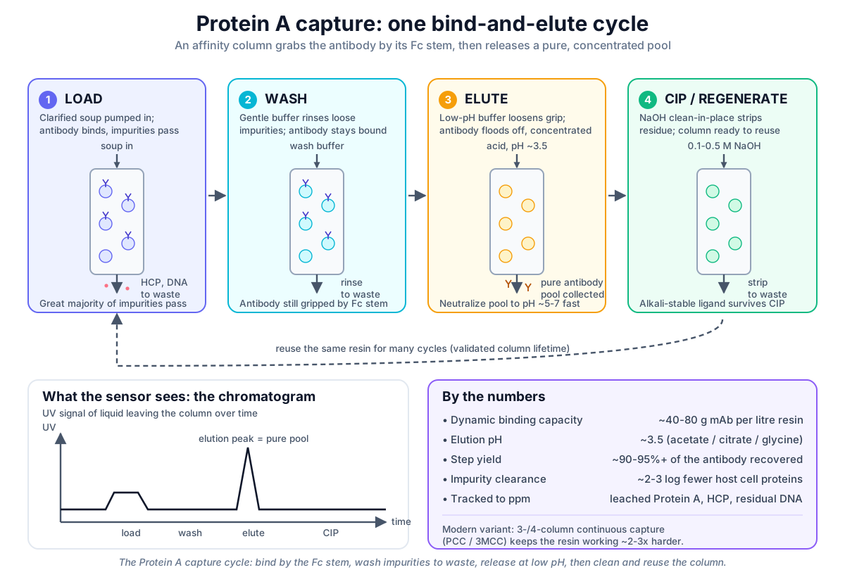 The Protein A capture cycle: bind the antibody by its Fc stem, wash impurities to waste, release a concentrated pool at low pH, then clean and reuse the column.
Original diagram by the authors, created with AI assistance.
The Protein A capture cycle: bind the antibody by its Fc stem, wash impurities to waste, release a concentrated pool at low pH, then clean and reuse the column.
Original diagram by the authors, created with AI assistance.

The column, field by field
Anatomy of a Protein A column, field by field
The first figure showed the cycle; the next zooms into the column as a documented object and the trace it draws. The column is not just "a tube of beads." It is a defined, documented piece of equipment with an identity card: a particular ligand, a packed-bed volume and height (the "bed" is the settled mass of beads inside the column — its volume and how tall it stands), a binding capacity, a residence time, and a proven packing quality. And in one cycle it produces a second object — a chromatogram with a pooling window — that is just as defined. The figure below lays out both: the column's fields on top, and the trace it draws on the bottom, with the slice we keep marked as the pooling window.
 A capture column reads as an identity card plus the chromatogram it produces: the ligand and bed define what it can hold, while the trace and its pooling window define what we actually keep.
Original diagram by the authors, created with AI assistance.
A capture column reads as an identity card plus the chromatogram it produces: the ligand and bed define what it can hold, while the trace and its pooling window define what we actually keep.
Original diagram by the authors, created with AI assistance.
The chromatogram's UV280 baseline (a sensor shining ultraviolet light at a wavelength of 280 nanometres, the band where proteins absorb most strongly, so the trace rises in step with how much protein is flowing past) stays low and flat through load and wash; if you push too much antibody on, a breakthrough shoulder rises as unbound product begins escaping to waste. At elution the trace jumps to a tall peak measured in milli-absorbance units (mAU, simply the unit on that UV signal's scale), and the operator collects only between two cut points — the pooling window — centred near a defined mark in column volumes (one column volume is the liquid volume the packed bed holds, the natural yardstick for measuring how much buffer has flowed through); we return to how operators choose these cut points under Failure modes below. Everything about this composite object (the fields on top, the cut points below) eventually becomes a structured record; the open-source build of that downstream chromatography step, including how a pooling decision is captured, is laid out in downstream chromatography. Those fields are not just a list — they are typed relationships a data model can read literally: this chromatogram is derived-from this column, the column is-a alkali-stable Protein A column that runs-on this PCC system, and the pool is derived-from a particular elution. Naming those edges explicitly is what lets a later system trace a finished vial back to the exact resin lot and chromatogram that made it; the ontology book builds that typed lineage in relations and genealogy and the class hierarchy behind it in classes and taxonomy.
Dynamic binding capacity, purity, and recovery
The headline number for any capture resin is its dynamic binding capacity (DBC) — how many grams of antibody one litre of packed beads can hold at the flow rate we actually run. We always quote it "at the running flow rate" because binding is mass-transfer (pore-diffusion) limited — meaning the bottleneck is simply how fast the antibody can travel into the bead's pores to reach a grabber: faster flow means a shorter residence time, which means a lower effective DBC, because the antibody cannot diffuse deep into the bead before the liquid sweeps past. (This is exactly what newer resins compete on — bead and pore designs that let the antibody diffuse in faster.) Modern Protein A resins typically reach a DBC of about 40 to 80 grams of antibody per litre of resin. That figure decides everything about column size: if your harvest contains, say, several kilograms of antibody, the DBC tells you how big a column you need (or how many load cycles a smaller column must run). Suppose the clarified harvest holds 3 kg of antibody and the resin's 50 g/L DBC is de-rated to a 40 g/L load with margin (de-rated meaning we deliberately load below the rated capacity for a safety cushion); then 3000 g divided by 40 g/L is 75 L of resin — or a smaller 25 L column run three times.
What does one pass actually buy you? Two things, mostly. First, purity: a single Protein A step removes roughly 2 to 3 orders of magnitude — "logs" — of host cell protein. (This is why the old shorthand "about 99 percent of impurities don't stick" is a friendly oversimplification: it is not that 99 percent of all impurity mass vanishes, but that HCP drops by 2 to 3 log, a hundred- to thousand-fold cut, in one move.) Second, recovery: a well-run capture step typically returns 90 to 95 percent or more of the antibody you loaded — very little medicine is lost. And because the antibody comes off in a small acid volume, the product stream is also concentrated several-fold compared with the dilute harvest that went in. Higher purity, high yield, smaller volume — all at once. That trifecta is why capture is the workhorse of the whole downstream train.
Why it matters
This one step does the heavy lifting of the entire purification. In a single pass it removes the vast majority of impurities, recovers nearly all the antibody, and shrinks the volume — and everything after this is fine-tuning by comparison.
Get it wrong and the consequences ripple downstream. Overload the resin and antibody breaks through to waste — lost medicine, lost money. Run the wash or elution poorly and impurities tag along; leftover host cell proteins could later provoke an immune reaction in a patient. There is also a sneaky impurity born in this very step: leached Protein A. A little of the grabber molecule itself can shed off the beads and ride along in the elution pool, so it becomes a tracked contaminant in its own right, controlled down to parts-per-million (nanograms of Protein A per milligram of antibody).
The purification step itself creates a new impurity. The very molecule we use to grab the antibody sheds a little of itself into the product — so the cleanup tool becomes something the cleanup must later remove.
Because patients inject this medicine straight into the body, purity here is a direct safety question, not a nicety. Capture is also the first place we can finally read, on the chromatogram, how much antibody we truly made.
The hardware: resin and column
The resin: engineered alkali stability and reuse durability
Protein A resin lives a hard life. Between every cycle it is washed with sodium hydroxide — typically 0.1 to 0.5 M (molar) NaOH, all three names (sodium hydroxide, caustic, NaOH) being the same strong alkaline cleaner — to strip away any clinging impurities and kill stray microbes, so the same beads can be reused for dozens or even more than a hundred cycles. The problem is that natural Protein A, being a protein itself, falls apart under that much caustic. So the industry engineered tougher versions: the binding domains of Protein A were redesigned — proteins are built from a chain of small building blocks called amino acids, and one of them, asparagine, is the spot most easily damaged by alkali, so swapping it out removes the weak link — and the redesigned ligand survives repeated caustic cleaning without losing its grip [3]. These engineered, alkali-stable ligands (often multimers — several copies stacked together — of a single redesigned binding domain, known as the B- or Z-domain) are what make modern resins both durable and cheap to run per gram of product.
You will meet these resins by brand name in any real facility. Cytiva's MabSelect SuRe and SuRe pcc, Purolite/Repligen's Praesto Jetted A50, Thermo Fisher's POROS MabCapture A, Merck's Eshmuno A, and JSR's Amsphere A3 are all alkali-stabilized Protein A resins competing on capacity, durability, and the speed at which the antibody can diffuse into the beads. They are not cheap: Protein A resin is one of the most expensive single materials in the plant, commonly running into the thousands of US dollars per litre — which is precisely why engineers care so much about squeezing the most product out of every litre, and why the economics push hard toward intensification [7].
Packing and proving the column: HETP, asymmetry, qualification
A column is only as good as how evenly its beads are packed. Channels or voids in the bed let liquid race through unevenly, smearing the elution peak and dragging impurities along. So before a column is trusted, it is qualified by injecting a harmless tracer (a small pulse of salt or acetone) and measuring how sharp and symmetric the resulting peak is. Two standard numbers come out: HETP (height equivalent to a theoretical plate — a "plate" is an imaginary thin slice of the bed that does one notional round of separation, so a bed of many short plates separates more sharply; smaller HETP therefore means a tighter, more efficient bed) and peak asymmetry (how lopsided the peak is — close to 1.0 is ideal). Typical capture columns run bed heights of about 10 to 25 cm.
Facilities choose between self-packed columns — large stainless or glass hardware the site packs and re-packs itself — and single-use, pre-packed columns that arrive ready to plumb in and are thrown away after a campaign, such as Cytiva's ReadyToProcess line or Sartorius's pre-packed formats. Single-use saves cleaning validation and cross-contamination worries; self-packed wins on cost per gram at very large scale. Either way, the packing quality is checked and documented, because a poorly packed capture column quietly poisons every step that follows.
Critical decisions and failure modes
Capture is forgiving in routine operation and brutal when it goes wrong, because it sits upstream of everything. Three failure modes are worth knowing by name.
Overloaded resin and breakthrough loss. Push more antibody onto the column than its dynamic binding capacity can hold at the running flow rate, and the excess simply does not bind — it breaks through and flows out to waste. On the chromatogram this shows as the breakthrough shoulder climbing during load: a quiet, expensive leak of product. The defence is to load to a fraction of DBC (with margin) and to watch the load trace, which is exactly why continuous multi-column capture catches the breakthrough from a nearly full column on the next column in line rather than losing it [1]. This is also where the field is starting to add a predictive layer: because breakthrough and the right pooling cut points show up as patterns in the UV trace, a soft sensor (a model that infers a hard-to-measure quantity from easier signals, here estimating the column's remaining capacity or the cleanest cut point in real time) can flag an approaching breakthrough before it shows on the chromatogram and steer the pooling window batch-to-batch. The ML book works exactly this step in capture chromatography models.
A misplaced pooling window. The operator collects only the slice of the elution peak between two cut points. Set those cut points too wide or too late and the tail of the peak — richer in aggregates, leached ligand, and trailing impurities — drags into the pool; set them too narrow and good product is sent to waste. Where the cut points fall, and against what UV thresholds, is a real-time quality decision, and it must be recorded as a reviewable part of the batch record. Chromatography data integrity — that the raw trace, the integration, and the pooling decision are all complete, attributable, and unaltered — is a recurring inspection finding, and guidance such as PIC/S PI 041 (PIC/S — the international Pharmaceutical Inspection Co-operation Scheme, which aligns drug-inspection agencies across countries; PI 041 is its data-integrity guide) on data management and integrity in GMP environments frames how those electronic records and decisions must be controlled [9]. The same expectation has a name: the chromatogram and its pooling record must be ALCOA+ — Attributable, Legible, Contemporaneous, Original, and Accurate, plus complete, consistent, enduring, and available — and the chromatography software that captures them is itself validated and access-controlled (electronic-signature and audit-trail rules, often cited as 21 CFR Part 11 or its EU twin Annex 11). Modern practice is shifting from exhaustive scripted computer system validation (CSV) toward risk-based computer software assurance (CSA), which concentrates testing on the highest-risk functions — here, the pooling-decision and audit-trail logic. The sister data book treats these in full: data integrity and ALCOA+ and the move from CSV to CSA.
Leached Protein A as a tracked contaminant. No ligand is perfectly anchored; a little Protein A always sheds off the beads and rides along in the elution pool. Leaching climbs with harsher elution, resin age, and proteolytic activity in the feed (the presence of protein-chopping enzymes that nibble the ligand), and the amount creeps up as the resin is repeatedly cleaned. Because leached Protein A is itself an impurity that can provoke an immune response, it is specified and measured for every batch — alongside host cell protein, or HCP (quantified in parts-per-million by ELISA, a lab test that uses tailored antibodies to detect and count a specific protein), and residual host-cell DNA (in nanograms per dose by qPCR, a test that amplifies and counts DNA) — under ICH Q6B (ICH — the International Council for Harmonisation, which sets the global drug-quality guidelines that regulators adopt), with acceptance limits in the parts-per-million range (nanograms of Protein A per milligram of antibody) [4]. A rising leached-ligand trend is one of the signals that a column has reached the end of its validated lifetime. The good news is that the very next steps help here too: capture alone often cannot push leached ligand under the spec, but a downstream polish (a later, finer purification step — the polishing chromatography ion-exchange step, which separates molecules by their electrical charge instead of by shape) reliably drives it down to single-digit or sub-ppm levels, and the low-pH hold of viral inactivation adds further aggregate and impurity clearance — so capture does not have to be perfect on its own.
In the real world
The standard commercial recipe is a Protein A platform process — "platform" meaning a single standardized, reusable recipe applied across many different antibody products with only minor tweaks: one column, run as a batch — load, wash, elute, clean, repeat — the same backbone across most approved mAbs [1]. It is reliable, well understood by regulators, and used industry-wide — run, like every step in this book, under current Good Manufacturing Practice (cGMP), the enforceable rules for making medicine safely and consistently. Its weakness is arithmetic: with a single column, the expensive resin sits idle during washing, elution, and cleaning, so a lot of costly material does nothing for much of each cycle.
The modern approach links several small columns together so capture never stops. The general industry term is periodic counter-current (PCC) chromatography — "counter-current" because the columns are arranged so the partially-used loading liquid from one column is fed onward to catch product on the next, the feed and the columns advancing against each other in a staggered relay — and you will also hear it called multi-column or sequential multi-column capture — the older "3MCC" label simply meant a three-column version of this idea. The trick: while one column is being washed, eluted, or cleaned, the next column is already loading, and the breakthrough from a nearly full column is caught by the next one in line so almost no antibody escapes. Commercial systems include Cytiva's ÄKTA pcc, Sartorius's Resolute BioSC, and Pall's Cadence BioSMB. Running this way raises resin productivity roughly two- to three-fold and cuts buffer consumption substantially, because each litre of expensive resin is working almost all the time rather than waiting [5].
Continuous capture shines brightest when it is fed continuously. Pair a multi-column capture skid with a perfusion bioreactor — a tank run in perfusion mode, continuously fed with fresh nutrients while spent liquid and product are drawn off, so it pours out a steady, gentle stream of harvest rather than one giant batch — and you have integrated continuous bioprocessing, where upstream and downstream flow together as one connected line instead of stop-and-go stages [6]. It is worth being precise here: fed-batch (the conventional mode where one tank is filled, fed top-ups of nutrient as it runs, then harvested all at once) with single-column Protein A remains the dominant approved process today. Continuous capture is the rising, not the reigning, approach.
There is one more reason this step is so closely watched: the rulebook. Under the international guideline ICH Q6B, the captured product's quality attributes — leached Protein A, host cell protein (measured by an ELISA assay), and residual host-cell DNA — are formally specified, with acceptance limits a batch must meet to be released [4]. Capture is also validated for the unglamorous but vital details: that nothing carries over from one batch to the next, that the resin still performs after being reused many times, and that a defined column lifetime is not exceeded. Done right, the very same beads safely make medicine again and again.
Finally, capture hands off neatly to the next safeguard. That low-pH elution pool is not just convenient chemistry — it is the opening move in a deliberate, orthogonal viral-clearance strategy — "orthogonal" meaning the layered steps work by different, independent mechanisms, so a virus that slips through one is caught by another. Holding the antibody briefly at low pH inactivates many enveloped viruses (viruses wrapped in a fatty outer membrane, which acid disrupts; non-enveloped viruses lack that membrane and so survive low pH, which is why it clears "many" but not all), which is exactly why the captured pool sets up the dedicated low-pH viral inactivation step that follows. Modern guidance such as ICH Q5A(R2) — the second revision (R2) of ICH's dedicated viral-safety guideline — frames how these layered, independent steps together assure viral safety [8]. Keeping bioburden low and cleaning thoroughly between cycles protects both yield and that safety margin.
Key terms
- Chromatography — separating a mixture by pumping it through a column where some molecules stick and others flow past.
- Column — the tube packed with beads that the liquid flows through.
- Resin — the millions of tiny porous beads packed inside the column.
- Protein A — a bacterial-derived grabber molecule, coated on the resin, that selectively binds IgG-type antibodies by their Fc stem.
- Affinity chromatography — chromatography that binds one specific target using a tailor-made grabber like Protein A.
- Buffer — a pH-controlled salt-water solution used to equilibrate, wash, elute, or re-equilibrate the column.
- Load / wash / elute — apply the soup, rinse off loose impurities, then release the captured antibody.
- Elution — using a low-pH buffer (around pH 3.5) to make the antibody let go, as a pure, concentrated stream.
- Chromatogram — the graph of what leaves the column over time; the tall, sharp peak is the pure antibody.
- Host cell protein (HCP) — stray protein from the factory cells, cut by roughly 2–3 log in a single capture step and controlled by ELISA.
- 3MCC — an older name for a three-column continuous capture system; the general term today is periodic counter-current (PCC) chromatography.
- Fc region — the bottom "stem" of the Y-shaped antibody that Protein A grips.
- Dynamic binding capacity (DBC) — how many grams of antibody one litre of resin can hold at the working flow rate (typically ~40–80 g/L).
- Residence time — how long the liquid lingers in the column (~3–6 minutes on load) so the antibody can diffuse in and bind.
- Leached Protein A — ligand that sheds off the beads into the product, tracked to parts-per-million levels.
- Clean-in-place (CIP) — scrubbing the column in place, usually with 0.1–0.5 M sodium hydroxide, so it can be reused.
- Pooling window — the slice of the elution peak, between two operator-set cut points, that is collected as product; everything outside it goes to waste.
- Breakthrough — antibody escaping unbound to waste once the resin is loaded past its capacity, seen as a rising shoulder on the load trace.
- HETP / peak asymmetry — the two qualification numbers from a tracer pulse: HETP (smaller is a tighter bed) and asymmetry (near 1.0 is ideal).
- Periodic counter-current (PCC) chromatography — multi-column continuous capture that keeps the resin working almost nonstop.
- cGMP — current Good Manufacturing Practice, the enforceable rules for making medicine safely and consistently.
- ALCOA+ — the data-integrity principle that a record (here the chromatogram and pooling decision) must be Attributable, Legible, Contemporaneous, Original, and Accurate, plus complete, consistent, enduring, and available.
- CSV / CSA — computer system validation (the older, exhaustive scripted approach) versus computer software assurance (the risk-based successor that focuses testing on the highest-risk functions).
- Soft sensor — a model that infers a hard-to-measure quantity (like remaining column capacity or the best pooling cut point) from easier signals such as the UV trace.
Where this leads
Our antibody is now a concentrated, low-pH pool — far purer than the soup we started with, and already sitting in acid. That is no accident. In the next chapter, viral inactivation, we take advantage of that low pH to neutralize any lurking viruses before we ever raise it back up — turning a quirk of the capture step into the first dedicated line of viral defense.