Building the factory cell
📍 Where we are: Stop 6 of 21 — we have the antibody's gene in hand, and now we turn ordinary living cells into tiny, reliable medicine factories.
In the last step we found the perfect antibody and read out its blueprint: a gene, a short stretch of DNA that holds the instructions for building one specific protein. But a gene is just a recipe on paper. To actually make the medicine, we need a living cell to read that recipe and churn out the protein, day after day, batch after batch, for years. This chapter is about building — and then carefully freezing — that cell.
Imagine you want the best bread in the world. You hand the same recipe to thousands of bakers, watch who bakes the most loaves of the highest quality, and crown a single champion. Then you make an army of identical copies of that one baker and freeze them. Every loaf you ever sell, for the next twenty years, comes from a thawed copy of that exact baker. That frozen army is your guarantee that today's bread tastes exactly like next decade's — and, crucially, that you can prove where every loaf came from.
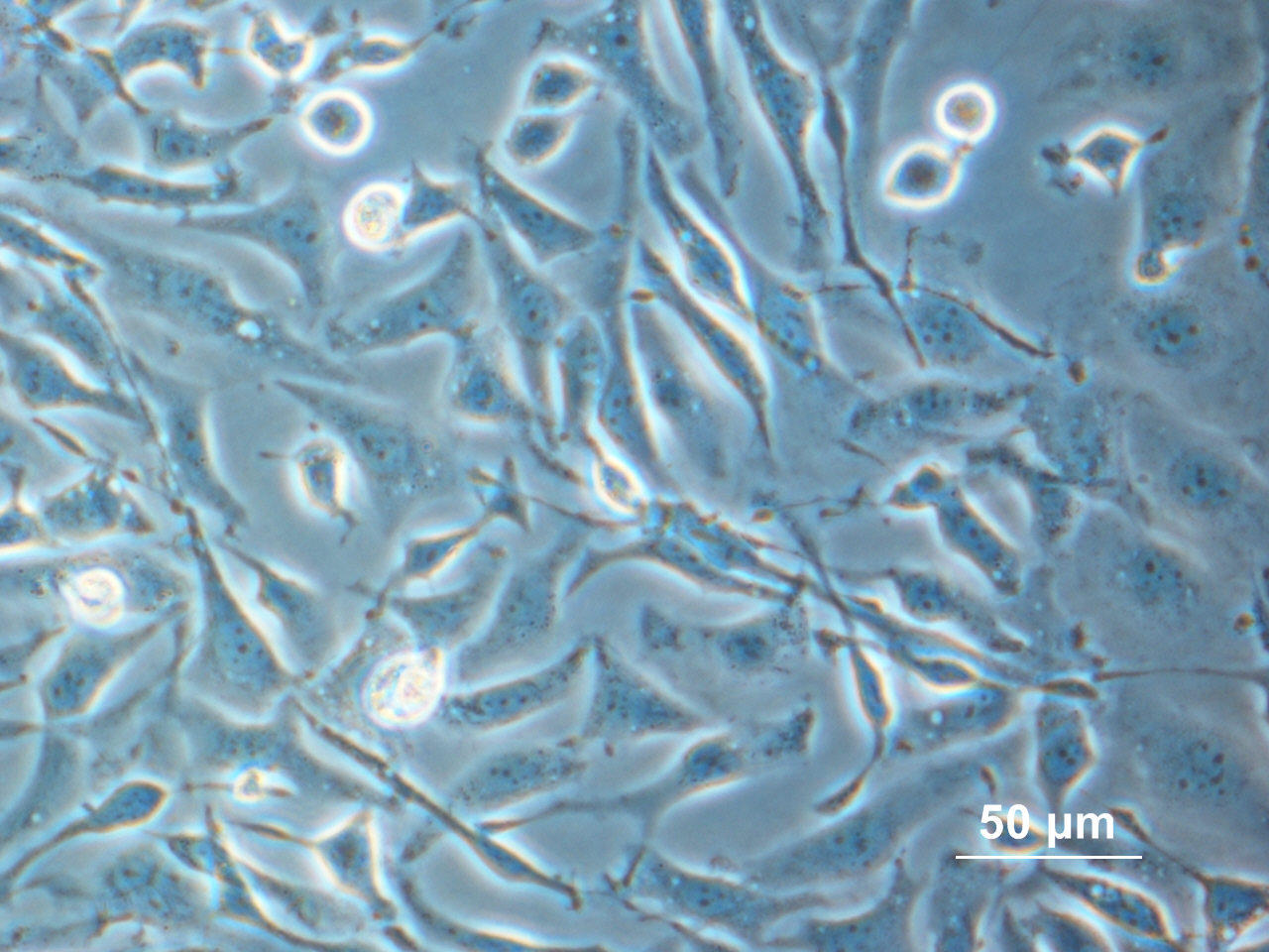 Adherent Chinese hamster ovary (CHO) cells viewed under a phase-contrast microscope — the workhorse mammalian cell line behind most therapeutic antibodies. The 50 µm scale bar shows how small they are.
Adherent CHO cells under phase-contrast microscopy. Image by Alcibiades, public domain, via Wikimedia Commons.
Adherent Chinese hamster ovary (CHO) cells viewed under a phase-contrast microscope — the workhorse mammalian cell line behind most therapeutic antibodies. The 50 µm scale bar shows how small they are.
Adherent CHO cells under phase-contrast microscopy. Image by Alcibiades, public domain, via Wikimedia Commons.
What this chapter covers
We will walk the whole journey from a gene to a frozen, regulator-approved seed stock. We meet the CHO cell and learn why it became the industry's favorite host. We see how the gene gets inside (transfection), how survival pressure weeds out the failures (selection), and how a single founder cell is hunted down and proven to be the one (cloning and screening). Then we build the two-tier vault every biologic depends on — the Master Cell Bank and the Working Cell Bank — and look at how they are frozen, tested, split between cities, and used (carefully) for decades. Throughout, we keep one question in view: why does so much of medicine safety trace back to this one cell?
Meet the host cell: CHO
The cell we use is almost always a CHO cell (Chinese Hamster Ovary cell) — a mammalian cell line first established in the 1950s and adapted over decades into the dominant workhorse of the biologics industry. The great majority of approved monoclonal-antibody therapeutics are made in CHO cells, and modern CHO cultures can secrete antibody at titers measured in grams per litre of broth — a thousandfold improvement over the milligram-per-litre yields of the early days [1]. (Titer is simply the amount of product the cells make per litre of culture.)
Why CHO and not, say, bacteria? Antibodies are large, folded proteins decorated with sugar chains — a pattern called glycosylation — and those sugars have to look human enough for the drug to work safely in patients. Bacteria cannot add human-like sugars at all; CHO cells can. CHO also grows well in suspension in large stainless-steel or single-use tanks, tolerates the chemistry of industrial culture, and — important for regulators — has a long, well-characterized safety history, including a known and manageable profile for viruses [6] — because any living cell line can carry hidden viruses, regulators need to know exactly which ones a host might harbour and that they can be detected and cleared (a concern we return to under adventitious agents). It is the "extensively studied, behaves predictably" cell, which is exactly what you want when the same cell will be making medicine for twenty years.
From gene to founder cell
Here is how a plain CHO cell becomes a dedicated antibody factory.
Transfection: getting the gene inside
Transfection just means delivering the antibody DNA into the cells so they can read it. In the lab, an operator mixes the purified antibody DNA with a small suspension of millions of CHO cells — in a cuvette for electroporation, or a culture vessel for lipofection — so the cells are exposed to the gene all at once, but most cells take up nothing. The two common methods trade off speed against gentleness. Electroporation zaps the cells with a brief electric pulse that opens temporary pores in the membrane, and commercial systems built for this — such as Lonza's Amaxa Nucleofector or Bio-Rad's Gene Pulser — can get the DNA into a large fraction of the surviving cells [2]. Lipofection instead wraps the DNA in tiny fat bubbles that fuse with the membrane; it is gentler to handle but typically less efficient and can be more toxic at the doses needed. Either way, a lucky minority of cells tuck the new gene into their own chromosomes, where it can be passed on to all their descendants.
Selection: survival of the gene-carriers
Now we apply pressure so that only cells that kept and are using the new gene can live. The trick is to deliver, alongside the antibody gene, a "survival gene" the cell needs — and then take away what that gene's protein produces. In the widely used glutamine synthetase (GS) system, selection runs in glutamine-free medium so that cells must make their own glutamine using the added GS enzyme — and, critically, the medium also carries the GS inhibitor methionine sulfoximine (MSX), which suppresses the cell's own (endogenous) GS so that only cells with high copies or strong expression of the transfected GS survive. Raising the MSX dose raises the stringency and can drive higher expression, directly paralleling the amplification step (forcing survivors to carry ever more copies of the gene) in the older system described next; modern GS hosts are typically GS-knockout (the host's own GS gene is deliberately removed) to sharpen the selection.
In that older methotrexate (MTX) / DHFR system (DHFR = dihydrofolate reductase, an enzyme the cell needs to grow), the drug methotrexate blocks DHFR, and only cells carrying many copies of the rescuing gene push through — turning up the antibody gene's copy number in the process [2]. Selection typically runs over 2–4 weeks and culls the population from millions of cells down to a much smaller field of survivors.
But surviving selection only proves the selection marker works — not that the antibody gene does. The two are separate risks: a survivor can keep an active survival gene while silencing, truncating, or losing the antibody gene that was meant to ride along with it. That is exactly why high-titer screening (not mere survival) is required, and why developers deliberately weaken the survival marker — for example by driving it from a feeble on-switch (a weak promoter, the stretch of DNA that controls how strongly a gene is turned on) or burying it inside a spliced-out spacer region (an intron, a non-coding piece cut out before the gene is read) — so that a cell can survive only if it integrated many active copies of the inserted DNA; and because the antibody gene rides along on that same DNA, the survivors are biased toward high antibody expression. Either way, some survivors make a trickle of antibody, some a flood, and some are unstable.
Cloning: finding the one founder
This is the crucial part, and the reason the chapter exists. We separate the cells so that each one sits alone, then let each single cell grow into its own small colony. A population grown from one isolated founder cell is a clone. To do this at industrial rigor, labs sort single cells into the wells of 96-well or 384-well plates, either by limiting dilution (diluting the cells so thin that, on average, each well receives one) or by flow-cytometry single-cell sorting, in which a machine deposits exactly one cell per well and often photographs it to prove it was alone. Modern automated imagers document each well at the moment of seeding, providing the assurance of clonality — visual evidence that the colony grew from a single cell — that regulators expect [3].
Screening: testing hundreds of candidates
A typical industrial campaign isolates and screens somewhere in the range of 500 to 5,000 clones (smaller programs screen fewer; high-throughput platforms more), winnowing them over roughly 3–6 months [2]. Each surviving candidate is judged on several axes at once: high titer, healthy growth rate, and — just as important — product quality. Titer alone can mislead, because titer (g/L) is roughly specific productivity (qP — the picograms each cell secretes per day) multiplied by the integral of viable cell density (IVCD) over the run — IVCD is simply the running total of how many live cells were present, summed across every day of the culture (a "cell-days" tally: more cells, alive for more days, equals more total product). A clone can reach the same titer by growing to a high cell density or by being highly productive per cell, and the two behave very differently at scale — which is why screening teams track qP directly, since a high-density but low-qP clone often disappoints at scale-up. Modern fed-batch CHO mAb platforms commonly reach about 2–8 g/L (high-performing platforms reaching ~10 g/L); early shake-flask or microbioreactor screens — typically just a few millilitres of culture, far smaller than the thousands of litres of a production tank — run lower and only rank-order the candidates, because tiny vessels cannot reproduce the fed-batch conditions that drive the highest titers. On the quality side the team names specific critical quality attributes (CQAs) — product properties that affect safety or efficacy — and ties each to its assay:
| Quality attribute | What it is | How it is measured |
|---|---|---|
| Aggregates | Clumped, misfolded antibody that can trigger an unwanted immune reaction | Size-exclusion HPLC (SEC), a lab method that sorts molecules by size |
| Charge variants | Small chemical changes (deamidation, C-terminal lysine, glycation) that shift the antibody's electrical charge | Imaged capillary isoelectric focusing (icIEF) or cation-exchange chromatography |
| Glycosylation pattern | The sugar chains on the antibody; e.g. afucosylation (removing a particular sugar, fucose) strengthens the antibody's ability to recruit immune cells to kill a target — antibody-dependent cell-mediated cytotoxicity (ADCC) — which is wanted for some cancer antibodies and unwanted for others, so it must be held in a consistent range | HILIC-fluorescence of the released sugar chains (glycans) |
(A CQA is a property of the product you must keep in range; a critical process parameter (CPP) is a knob on the process you set to keep CQAs in range — both are ICH Q8 (Quality by Design) concepts.) A clone that grows fast but makes subtly wrong protein is disqualified. The goal is a single founder that is high-producing and makes the right molecule and stays stable over many cell generations [4].
This whole campaign is a winnowing funnel: a wide crowd of candidates narrows, stage by stage, to one survivor that then seeds a strict bank hierarchy. That funnel is also a natural place for a predictive model — software that learns from past clones to forecast a new one's behaviour: instead of waiting months to see which high-titer clone stays stable at scale, teams increasingly train models on early imaging, growth-curve, and quality readouts to rank-order candidates and flag the ones likely to drift. The machine-learning book treats this clone-ranking problem in full in its cell-line development chapter.
 From a crowd of clones to one founder cell, then a two-tier vault: each stage winnows the field, and the lone survivor seeds the Master and Working Cell Banks.
Original diagram by the authors, created with AI assistance.
From a crowd of clones to one founder cell, then a two-tier vault: each stage winnows the field, and the lone survivor seeds the Master and Working Cell Banks.
Original diagram by the authors, created with AI assistance.
Every one of these screening readouts — the titer for each well, the aggregate percentage, the clonality image, the well-plate map — is also a data point with a birth, an owner, and a timestamp. How that flood of measurements is captured, contextualized, and made trustworthy is the subject of the data book's chapter on where data is born.

Why clonality is non-negotiable
That single champion clone becomes the cell line for this product. Because every cell descends from one founder, the population is clonal — and clonality is a regulatory expectation for production cell banks, not a nicety. International guidance, anchored by ICH Q5D (the harmonized standard for deriving and characterizing cell substrates), frames the cell bank as starting from a single clonal isolate with a fully documented history [5]. The industry's own consensus is that clonal derivation matters because it underpins consistency, while being honest that clonality is one element of a broader control strategy, not a magic guarantee of perfection [3].
A subtle but important truth: clonality at the start does not freeze the cell line in amber forever. CHO genomes are restless — over many divisions, clones can pick up mutations, silence the inserted gene, or drift in productivity [4]. That is precisely why we also confirm, using the framework of ICH Q5B, that the correct antibody coding sequence is integrated (stitched into the cell's own chromosomes, as in transfection above) and stays intact from the cell bank through to the end of production [9]. Clonality gives you a clean, single-origin starting point; ongoing characterization is what keeps it trustworthy.
When the founder drifts: passage instability and silent contamination
The most expensive failure in this whole chapter is also the quietest: a clone that looks perfect at the bank and slowly betrays you in production. A stable high-producer banked at, say, passage 5 can silence the inserted gene or shift its N-glycosylation pattern by passage 50 — not because anything broke, but because CHO genomes rearrange, methylate (add chemical tags that switch genes off without changing the DNA letters), and lose transgene copies (copies of the inserted antibody gene) over many divisions [4]. The titer sags, or the sugar profile drifts out of the comparability window (the acceptable range within which a batch still counts as "the same product" as before), and downstream batches fall out of specification — the documented acceptance limits a batch must meet to be released — even though the process never changed. This is exactly the drift that ICH Q5D, Section 3.2 anticipates when it requires a defined limit of in-vitro cell age — a passage ceiling demonstrated, not assumed, by growing the line past the production limit and proving identity, productivity, and product quality still hold [5].
A second silent failure mode is contamination that the assay says is not there. Mycoplasma — the tiny wall-less bacterium screened for at release — is notorious for evading detection: it can sit below the limit of a single test method, and a careless or single-method screen returns a false negative while the culture is in fact infected, subtly distorting growth and glycosylation for months [10]. The lesson both failure modes teach is the same: a clean release certificate is a snapshot, not a guarantee. Trust comes from the combination of a derivation history, a demonstrated passage limit, and orthogonal contamination testing — never from any one number alone. We will watch this passage clock again at the seed train, where the cell-age limit is enforced batch by batch.
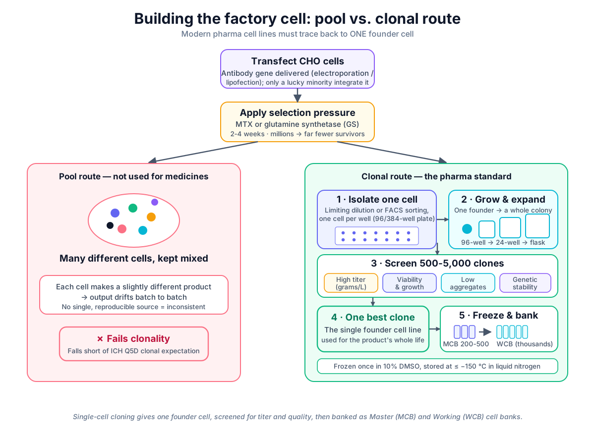 Single-cell cloning ensures clonality and consistency. Unlike early industrial pools, modern pharma cell lines originate from one founder cell, tested for high titer, low aggregates, and genetic stability before being frozen into Master (MCB) and Working (WCB) cell banks.
Original diagram by the authors, created with AI assistance.
Single-cell cloning ensures clonality and consistency. Unlike early industrial pools, modern pharma cell lines originate from one founder cell, tested for high titer, low aggregates, and genetic stability before being frozen into Master (MCB) and Working (WCB) cell banks.
Original diagram by the authors, created with AI assistance.
The two-tier vault: MCB and WCB
How the architecture works
Once we have our founder clone, we do not just keep it growing on a bench — a single contamination or freezer accident could erase the entire product. Instead we build a two-tier cell bank, the system every biologic medicine rests on [7].
Master Cell Bank (MCB). We expand the founder clone, then split that one uniform culture into a set of identical vials — typically a few hundred (often in the range of 200–500), each holding around 1–2 mL of cells at roughly 1–5 million viable cells per mL. The vials are frozen once, directly from this expanded founder culture, in a culture medium spiked with about 10% dimethyl sulfoxide (DMSO) — a cryoprotectant that keeps ice crystals from shredding the cells as they freeze. They are then stored in liquid nitrogen. Here is a point the friendly shorthand of "minus 196 °C" gets slightly wrong: vials held in the vapor phase above the liquid nitrogen sit at roughly −150 °C to −180 °C, while vials submerged in the liquid phase reach about −196 °C. Both are allowed by regulators; vapor-phase storage is very common because it avoids the cross-contamination risk of shared liquid, while liquid-phase storage runs colder and is sometimes preferred for the longest-term holds. The shared point is simply that life is paused. This frozen set — the MCB — is the irreplaceable seed stock, the single source from which everything downstream descends.
Working Cell Bank (WCB). We do not reach into the precious MCB for everyday work. Instead we thaw one MCB vial, grow it for a short, defined amount of cell age — measured not in calendar time but in how many times the cells divide (on the order of 5–10 population doublings) — and then freeze new vials from that culture. (A note on units: a passage is one subculture or split, while a population doubling is one doubling of biomass — several doublings usually occur per passage. The limit of in-vitro cell age is most rigorously expressed in population doublings (PDL) or generations; "passage number" is a coarser proxy. We keep the WCB and table in doublings for that reason.) This is the WCB, and a single MCB vial can seed thousands of WCB vials (often in the 2,000–5,000 range). Each WCB vial is, again, frozen once. The WCB is the working supply: every production batch starts by thawing one WCB vial. The hierarchy is strict and one-directional — the founder clone makes the MCB, the MCB makes the WCB, and only the WCB touches routine manufacturing [7]. When a WCB runs low, you thaw another MCB vial and make a fresh WCB; you do not re-clone or re-derive.
The two tiers side by side:
| Attribute | Master Cell Bank (MCB) | Working Cell Bank (WCB) |
|---|---|---|
| Made from | The single founder clone, expanded | One thawed MCB vial, grown ~5–10 doublings |
| Vials in the set | ~200–500 | ~2,000–5,000 |
| Each vial | ~1–2 mL at ~1–5 million cells/mL, frozen once | Frozen once |
| Frozen and stored in | ~10% DMSO, in liquid nitrogen (−150 to −180 °C vapor, or −196 °C liquid) | Same medium and storage |
| Role | Irreplaceable seed stock — never used for routine production | Working supply — every production batch thaws one vial |
Anatomy of one MCB vial
It is tempting to picture an MCB vial as just a tube of frozen cells. It is not. Each vial is an identity card: a tightly defined object whose every attribute — how much liquid, how cold, how many cells, which tests passed — is recorded and is the thing every future batch traces back to. Reading one vial top to bottom shows what a cell bank actually is.
 One MCB vial as an identity card: physical contents on top, the release tests that gate the bank in the green core, and its one-directional lineage to founder, WCB, and every batch in the violet panel.
Original diagram by the authors, created with AI assistance.
One MCB vial as an identity card: physical contents on top, the release tests that gate the bank in the green core, and its one-directional lineage to founder, WCB, and every batch in the violet panel.
Original diagram by the authors, created with AI assistance.
The physical contents are modest — roughly 1–2 mL of culture at 1–5 million viable cells per mL, suspended in medium with about 10% DMSO — but the attached record is not. The viable-cell density (VCD) and viability at freeze are logged so that years later a technician knows what a healthy thaw should look like; the storage phase (vapor or liquid nitrogen) is recorded because it sets the exact temperature; and the founder passage number is captured because it starts the passage clock that the line must never outrun. The same identity-card thinking reappears across this trilogy: in the data book, the equivalent record is the structured data point with its own metadata (the who/when/units around it), and in the open-source companion the offline seed-train data is written as concrete rows you can query — see Open-Source Bioprocess Data Systems for that implementation.
Testing and regulatory release
Before either bank can be released, it is tested hard. Sterility and freedom from mycoplasma (a tiny, common contaminating bacterium), bacteria, and fungi are confirmed; the bank's identity is verified — that it really is CHO and really is this product's line — using methods like isoenzyme analysis (which reads the species-specific forms of certain enzymes to confirm the cells really are hamster, not a stray human or mouse line) or STR (short tandem repeat) profiling (a DNA fingerprint); and viral safety is evaluated for adventitious (uninvited) agents under ICH Q5A(R2) [6]. These characterization expectations are spelled out internationally by ICH Q5D [5] and WHO Technical Report Series 978 [7], the documents that directly govern cell substrates for therapeutic proteins. The FDA's Characterization and Qualification of Cell Substrates guidance [8] is written for cell substrates used in viral-vaccine production and is applied to therapeutic-protein banks by analogy; in the United States the binding rule is the cGMP requirement of 21 CFR Part 211 — where cGMP stands for current Good Manufacturing Practice, the rules for making medicines safely and consistently.
Why it matters
This one cell line is the source of everything that follows. Every future batch starts by thawing a single WCB vial, so the cell line's quality is literally baked into the medicine forever. Pick a clone that drifts or makes subtly wrong protein, and you have built a flaw into every dose for decades. Choose well, document everything, and you have a stable foundation that can outlast the careers of the scientists who built it.
Clonality and the cell banks also give traceability: any vial a patient's medicine ultimately came from can be traced straight back to these frozen banks, and from there to the single founder cell and its documented history. If a question arises years later — a quality signal, an audit, a regulatory query — the manufacturer can point to the exact bank, the exact founder, the exact records [5]. That is a backbone of patient safety: it is why batch number 5,000 can be trusted to be as safe and effective as batch number one. Notice that this one-directional chain — founder → MCB → WCB → batch — is a typed "derived-from" relationship, and the bank tiers themselves are is-a kinds of cell bank; a computer can follow such typed links automatically once they are written down as a formal model. That is exactly what the ontology book does, turning this lineage into a walkable graph in its chapters on classes and taxonomy and relations and genealogy.
In the real world
Because building a great cell line takes the better part of a year and tens of millions of dollars, companies guard their banks like crown jewels. A standard practice is geographic redundancy: the MCB is physically split, with the primary vials in one facility's liquid-nitrogen freezers and backup vials in a freezer in a different building, a different city, or a contracted third-party storage site — so a single freezer failure, fire, or flood cannot erase the line. These holdings follow documented rotation and monitoring schedules, and retained backup vials are periodically pulled and thaw-tested (often every 3–5 years) to confirm the cells are still viable and stable after years in the cold.
How long can one bank serve? In practice, a single MCB can support a product for its commercial lifetime — often two decades or more — but not by being grown forever. The line is used only up to a defined maximum passage number (commonly capped on the order of tens of passages, with cell lines often demonstrated stable to roughly 50–100 passages in production studies), beyond which it must be re-derived from a fresh MCB vial. Regulators increasingly emphasize characterizing this genetic and epigenetic drift over time and pinning down that limit-of-in-vitro-cell-age, consistent with the cell-substrate and expression-construct (the engineered package of DNA — the antibody gene plus its control elements — inserted into the cell) expectations of ICH Q5D [5] and ICH Q5B [9] [4]. So "used for decades" is true at the level of the bank, with each individual culture run kept young and within limits.
A trustworthy cell line is the upstream prerequisite for everything that follows: you cannot demonstrate a better process until you have a dependable cell making the product. The cell line built here will next feed the seed train and the production bioreactor, whether the plant runs the standard fed-batch process or a modern perfusion (continuous) process. The cell line comes first; the choice of how to grow it comes after.
Key terms
- CHO cell — Chinese Hamster Ovary cell; the dominant mammalian host used to manufacture antibody medicines, prized for human-like glycosylation and a long safety record.
- Transfection — getting the antibody gene inside a cell so the cell can read it, by electroporation or lipofection.
- Selection — applying survival pressure (e.g., GS or MTX systems) so only cells that kept and express the new gene live.
- Clone — a population of cells all descended from one single founder cell.
- Clonality — the property of a cell line coming from one founder; a regulatory expectation for production cell banks, supporting consistency.
- Cell line — the one chosen high-producing, stable clone used for a product, within defined passage limits.
- Titer — how much antibody a culture produces, typically measured in grams per litre.
- Glycosylation — the human-like sugar chains decorating an antibody; a key quality attribute CHO cells can produce and bacteria cannot.
- Aggregates / charge variants — quality defects (clumped protein; subtle chemical differences) screened out during clone selection.
- Cryoprotectant (DMSO) — a chemical (about 10% DMSO) added before freezing to stop ice crystals from killing the cells.
- Master Cell Bank (MCB) — the original set of identical frozen vials made once from the founder clone; the protected seed stock.
- Working Cell Bank (WCB) — vials made once from a thawed MCB vial and used to start everyday production batches.
- Maximum passage number — the defined ceiling on how many times a culture may be grown before re-deriving from a fresh MCB vial.
- Viable-cell density (VCD) — the count of live cells per millilitre, logged when an MCB or WCB vial is frozen so the expected healthy thaw is known.
- STR profiling — short-tandem-repeat fingerprinting used to confirm a cell bank's identity (that it really is CHO and really is this product's line).
- Limit of in-vitro cell age — the passage ceiling demonstrated under ICH Q5D, beyond which a culture must be re-derived from a fresh MCB vial.
- Traceability — the ability to trace any batch back to its exact source cell bank, founder cell, and documented records.
- Predictive model — software that learns from past clones to forecast a new one's behaviour, used to rank-order screening candidates before slow at-scale data is in.
- Typed lineage — the founder → MCB → WCB → batch chain written as explicit "derived-from" and "is-a" relationships a computer can follow, the raw material an ontology turns into a walkable graph.
In short
- One founder cell becomes the whole product: it seeds the Master Cell Bank, which seeds the Working Cell Bank, and only the WCB touches routine manufacturing.
- Clonality is necessary but not sufficient — it is the clean single-origin start, while ongoing characterization (identity, stability, viral and mycoplasma safety) is what keeps the line trustworthy.
- The bank outlives any single culture: each run is kept young within a demonstrated limit of in-vitro cell age, so "used for decades" holds for the bank, not for any one growing culture.
Where this leads
We now have the irreplaceable thing: a single, clonal, high-producing, fully documented cell line, frozen and split into Master and Working Cell Banks. But a brilliant cell is only as good as the conditions we grow it in. In the next chapter, Perfecting the recipe (process development), we leave the freezer and head to the lab bench to find the best recipe for this cell — the right food, temperature, oxygen, and sequence of steps that coax it to make the most high-quality medicine, reliably, at scale.