Finding the antibody
📍 Where we are: Stop 5 of 21 — we know our target, and now we must find the exact antibody that grabs it.
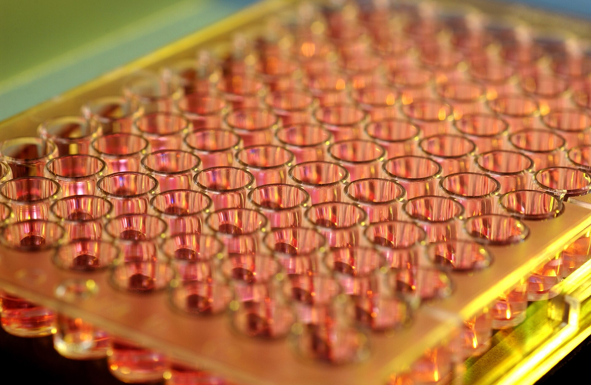 A multi-well microplate holds dozens of tiny cultures side by side, so a single screen can test many antibody candidates at once — the high-throughput sifting at the heart of discovery.
Multi-well microplate. Image by CSIRO, CC BY 3.0, via Wikimedia Commons.
A multi-well microplate holds dozens of tiny cultures side by side, so a single screen can test many antibody candidates at once — the high-throughput sifting at the heart of discovery.
Multi-well microplate. Image by CSIRO, CC BY 3.0, via Wikimedia Commons.
In the last step we picked a target — a specific molecule on or around a diseased cell that we want our medicine to grab. Now comes the treasure hunt: we have to discover the one antibody (a Y-shaped protein the immune system uses to recognize and stick to specific things) that binds that target perfectly. The prize at the end is not a vial of liquid. It is a piece of DNA — the exact instructions for building that antibody, the seed for every cell, batch, and dose that follows.
Think of the target as a lock. We need to find the one key that fits it. We can ask an animal's immune system to carve us a key, or we can rummage through a giant bin of billions of pre-made keys until one slides in. Either way, once we find the key that works, we file down its rough edges, make sure it will survive years in a pocket, and then write down its blueprint so we can stamp out perfect copies forever.
What this chapter covers
We will follow two parallel routes to a candidate antibody — the animal-based hybridoma method and the test-tube phage display method — and see the real numbers behind each: how long the mouse work takes, how big the phage libraries are, how many rounds of selection it takes to enrich the winners. Then we watch those candidates pass through three refining gates: affinity maturation (gripping harder), humanization (looking human enough to be tolerated), and developability (surviving the factory and the fridge). We will see why the modern industry increasingly skips the mouse entirely with fully human libraries and transgenic mice. Finally, we follow the survivors into a lead-selection funnel that weighs binding, kinetics, stability, and effector function together, and ends at a single, codon-optimized DNA sequence ready to hand to a cell — with a nod to the regulators who will want to know exactly where that sequence came from.
Anatomy of the antibody variable domains
Before we hunt for the antibody, it helps to know exactly what we are hunting for — and what the winner will have to carry with it for the rest of the journey. The business end of an antibody is its variable region, split across two short protein stretches: the heavy-chain variable region (VH) and the light-chain variable region (VL). Those two domains fold together to form the binding pocket, and everything this chapter does — selecting, maturing, humanizing, screening — is in service of shaping that pocket and the molecule around it.
But a lead candidate is never just "a sequence that binds." By the time it leaves this stage it is an identity card: a structure (VH + VL, plus six short gripping loops — the CDRs, complementarity-determining regions, detailed later under Humanization — sitting on a human framework) bundled together with a set of measured developability attributes — its affinity (Kd, a binding-strength number we explain below), its binding kinetics (kon/koff), its thermal stability (Tm), and its aggregation state — plus the Fc effector profile (Fc is the antibody's stable "tail," whose effector functions recruit the immune system — detailed later) and the final codon-optimized DNA (DNA rewritten to be read efficiently by the manufacturing cell, explained at the end of the chapter) that encodes it. Each of those numbers is born from a real instrument reading, and each will be re-measured again and again downstream. That structure-plus-attributes bundle is the thing every later chapter inherits.
 The lead candidate is an identity card, not a bare sequence: a VH + VL structure carrying its measured developability attributes (Kd, kon/koff, Tm, aggregation) forward to every downstream chapter.
Original diagram by the authors, created with AI assistance.
The lead candidate is an identity card, not a bare sequence: a VH + VL structure carrying its measured developability attributes (Kd, kon/koff, Tm, aggregation) forward to every downstream chapter.
Original diagram by the authors, created with AI assistance.
Notice the bottom panel of that card. The Tm, Kd, and aggregation readings do not just sit in a lab notebook — each one is generated by an instrument, recorded as a structured measurement, and stored in a system of record. In Data Management we follow how a single such reading becomes a governed data point (one with a known owner, timestamp, and audit trail), and in Open-Source Bioprocess Data Systems we see that same point land as a concrete row in a LIMS/ELN schema. The physical molecule is the anchor; the data thread starts here.
Two routes to a candidate
There are two classic ways to find a candidate antibody, and most modern programs run versions of both.
Route A — Ask an animal (the hybridoma method)
We inject a tiny, harmless amount of the target — the antigen — into a mouse, usually an inbred strain such as BALB/c, mixed with an adjuvant (an immune "alarm bell," classically Freund's adjuvant) that tells the immune system to take the intruder seriously. Over about 8 to 12 weeks of repeated injections, the mouse mounts what is called a polyclonal response: many different B cells (the immune system's antibody-making white blood cells) each wake up and start producing their own single kind of antibody. (It is worth being precise here. People sometimes say the mouse "makes millions of antibodies," but each individual B cell makes exactly one clonotype — one antibody sequence. The mouse as a whole produces a diverse mixture, and our job is to fish out the rare individual B cells whose one antibody grips our target best.)
The problem is that B cells, left alone, die after a few divisions. The Nobel-winning trick of Köhler and Milstein was to fuse those antibody-producing B cells with myeloma cells — cancerous bone-marrow cells that divide tirelessly — using polyethylene glycol (PEG) to melt the two membranes together [1]. Common myeloma fusion partners are non-secreting lines such as SP2/0-Ag14 or P3X63Ag8.653, chosen because they no longer make antibodies of their own to contaminate the result. Each fused cell, a hybridoma, inherits the B cell's single antibody recipe and the myeloma's stamina, and grows into a tiny factory churning out one pure antibody.
A caution that matters downstream: hybridomas are robust but not truly immortal in the "lives and divides forever" sense. Over many passages (rounds of growing and splitting the culture) they can lose chromosomes, stop secreting, or senesce (grow old and stop dividing). That fragility is one of the reasons the industry freezes a definitive Master Cell Bank early and works from controlled stocks rather than an open-ended bench culture — a theme we will pick up in the next chapter.
Route B — Search a library (phage display)
Instead of an animal, we can build a vast collection of antibody fragments in the laboratory and let the target itself pick the winners. Each fragment is displayed on the surface of a bacteriophage — a harmless virus that infects only bacteria, used here as a tiny billboard. The founding demonstration by McCafferty and colleagues showed that antibody variable domains could be stitched onto filamentous phage (commonly the M13 or fd phages) so that each phage carries its antibody on the outside and the gene encoding that same antibody on the inside [2]. The displayed fragment is usually a compact engineered piece — a single-chain variable fragment (scFv), where the two binding domains are tethered by a short linker, or a slightly larger Fab fragment.
A single library can hold roughly 6 to 10 billion different variants, with binding strengths spread across an enormous range. Because this strength is a dissociation constant (Kd) measured in M (molar, a concentration unit — moles per litre), a smaller number means a tighter grip — counter-intuitive at first, and we unpack why in the next section. So the spread runs from weak grips around 10⁻⁶ M down to very tight ones near 10⁻¹² M [5]. To enrich the good binders we run biopanning: we pour the library over the immobilized target (binding), rinse away everything that does not stick (washing), pry loose what remains (elution), and re-grow it in bacteria (amplification). Each cycle concentrates the binders; after 3 to 5 rounds the population shifts from a near-random sea to a small set of genuine winners. Phage display is not a museum piece — it is the discovery engine behind several approved therapeutic antibodies, the most famous being the blockbuster adalimumab [5].
Both routes are deliberately wasteful at the start, because finding a great antibody is a numbers game: genuine binders are rare — a handful out of the millions to billions of cells or clones examined — which is exactly why we begin with so vast a starting pool. (The exact "hit rate" is not a single number: it depends on the route and on what you count — positive hybridoma wells, enriched phage clones, or sequenced B cells all have very different denominators.)
Refining the candidates
A raw hit is rarely good enough to put in a person. Three gates turn a promising binder into a drug-grade molecule.
Affinity maturation — gripping harder, and the off-target trade-off
Affinity is how tightly an antibody holds its target, summarized by the dissociation constant (Kd): the smaller the Kd, the tighter the grip. Lab techniques such as error-prone PCR (deliberately copying the gene sloppily to sprinkle in mutations) or yeast display (the same select-and-enrich logic as phage, run on yeast cells) can improve a lead's affinity 10- to 100-fold, often into the single-digit-nanomolar or sub-nanomolar (nM; nanomolar is 10⁻⁹ M, the middle of the range introduced above) range, which is sufficient for most therapeutic mechanisms — though, as the next paragraph argues, tighter is not automatically better [6]. But "tighter is always better" is a myth worth puncturing. Pushing into the sub-nanomolar range can backfire: the molecule may start sticking to off-target look-alikes, or the extra mutations may hurt stability. Past a point, more affinity buys nothing and may cost developability.
Humanization — CDR grafting and framework preservation
A mouse antibody looks foreign to the human immune system, which may attack it and neutralize the drug. The fix is humanization, and it is more surgical than the shorthand "keep only the tiny gripping part" suggests. As we saw on the identity card above, an antibody's variable region has six short loops called complementarity-determining regions (CDRs) that do the actual gripping, sitting on a scaffold of framework regions. Classic CDR grafting, pioneered by Jones and colleagues, transplants all six mouse CDR loops onto a human framework [3]. But a naked graft often loses affinity, because some neighboring framework residues subtly shape the loops. So, as Queen and colleagues showed, humanization also keeps a handful of carefully chosen mouse framework residues to preserve the binding geometry while still replacing the large majority of the sequence with human germline (the standard human antibody gene templates the body inherits and treats as its own) [4]. The result looks human enough to be tolerated yet grips like the original.
Developability screening — Tm, DLS/SEC-HPLC aggregation, viscosity
A molecule can be a brilliant binder and still be undruggable if it clumps, gels, or falls apart. Developability is the catch-all for "can we actually make and store this?" Teams run a battery of biophysical screens early: differential scanning fluorimetry (DSF), also called a thermal-shift assay, measures the melting temperature (Tm) at which the protein unfolds (a Fab Tm above roughly 60 °C is preferred); dynamic light scattering (DLS) and size-exclusion HPLC (SEC-HPLC) flag aggregation and clumping; and viscosity is checked because subcutaneous drugs must stay injectable even when concentrated past 50 mg/mL.
Developability is not only biophysics: teams also scrub the sequence itself for liabilities. A liability is a chemically fragile spot in the protein's own sequence — a place where the molecule can slowly react and change its own chemistry over time (a sugar getting attached, an amino acid losing or rearranging an atom, or oxidizing) and so drift from one batch to the next. The usual suspects are N-linked glycosylation sites lurking in a CDR (the NxS/T motif), deamidation-prone (NG/NS) and isomerization-prone (DG) motifs, oxidation-prone Met/Trp residues, free cysteines, and N-terminal pyroglutamate — the parenthetical letter-codes are the residue patterns an expert scans for. These are designed out here precisely because, left in, they surface later as critical quality attributes (CQAs) — charge variants (slightly altered copies that carry a different electric charge) and fragments the factory must then monitor and control. Leads are also screened for polyreactivity / nonspecific binding (for example by baculovirus-particle ELISA or cross-interaction chromatography), because a polyreactive antibody clears fast and doses poorly no matter how tightly it grips its target. A large benchmark study of clinical-stage antibodies confirmed that real approved molecules cluster within "well-behaved" ranges on exactly these axes — and that flagging the badly behaved ones early saves enormous downstream pain [7]. The very same biophysical measurements reappear, more formally, when the molecule reaches Analytical methods and formulation as release-and-characterization assays — developability screening is just their early, exploratory rehearsal.
Increasingly, these wet-lab screens have an in-silico twin: a sequence-based model can flag the same liabilities and predict aggregation, viscosity, and immunogenicity straight from the VH/VL sequence, before a single clone is made. The Machine Learning & AI book works through how such a developability predictor is built and validated against exactly these readings.
When affinity maturation backfires
It is worth dwelling on the failure mode, because it shapes how good teams run the previous three gates. The intuitive program — "push affinity as hard as the platform allows, then worry about everything else" — is exactly the trap. Driving a phage- or yeast-selected variant to ever-tighter binding piles mutations into the CDRs, and those same mutations can destabilize the fold (a lower Tm) or broaden specificity so the antibody begins binding off-target look-alikes — a route to poor pharmacokinetics and, in some cases, immunogenicity in vivo [10]. The trade-off is structural: the residue changes that tighten the grip are often the ones that perturb the framework or expose hydrophobic patches that seed aggregation. This is why a sub-nanomolar binder with a 54 °C melting temperature loses to a slightly weaker binder that holds its shape — the theme of the decision matrix below. Modern affinity-maturation campaigns therefore co-screen stability and specificity alongside affinity from the start, rather than rescuing developability after the fact [10].

The lead-selection decision matrix
With a handful of refined candidates, the team must crown a single lead — the front-runner that will absorb years of development. This is not a single beauty contest but a weighted decision matrix that scores each candidate on several axes at once.
 The lead is chosen by a weighted matrix, not a single number: clone C2 has the tightest Kd but fails on stability and aggregation, so the balanced clone C4 — slightly weaker but manufacturable — wins.
Original diagram by the authors, created with AI assistance.
The lead is chosen by a weighted matrix, not a single number: clone C2 has the tightest Kd but fails on stability and aggregation, so the balanced clone C4 — slightly weaker but manufacturable — wins.
Original diagram by the authors, created with AI assistance.
The matrix makes the affinity-maturation trade-off concrete. Clone C2 would win any contest judged on Kd alone, yet its 54 °C Tm and 82% monomer fraction flag it as the molecule most likely to aggregate in a vial — so it loses to C4, which gives up a hair of binding for a structure that survives the factory and the fridge. Every column of this matrix — Kd, kon/koff, Tm, %monomer — is re-run, more rigorously, as a formal release assay once the molecule reaches Analytical methods and formulation; the screen here is the early, exploratory rehearsal.
Affinity alone is too crude, because how an antibody binds matters as much as how tightly. Binding has two speeds: the on-rate (kon), how fast the antibody finds and latches onto its target, and the off-rate (koff), how slowly it lets go. These are read directly off the association/dissociation curves of a label-free biosensor — surface plasmon resonance (SPR, e.g., Biacore) or bio-layer interferometry (BLI, e.g., Octet) — which is the standard tool for ranking clones against the very decision matrix above. Common goals are a kon in the 10⁵–10⁶ M⁻¹s⁻¹ range and a koff of 10⁻⁴ s⁻¹ or slower, depending on the target and the residence time the mechanism needs — these are context-dependent goals, not fixed pass/fail specs. In-vitro potency is measured in functional assays (reported as an IC50, the concentration that produces half the maximal effect, using readouts such as AlphaScreen or ELISA), and thermostability is folded back in through the melting temperature (Tm) from the developability screens [6].
Crucially, real-world effectiveness depends on more than the binding pocket. An antibody's lifetime in the bloodstream — its serum half-life — determines how often a patient must be dosed, and is tuned largely by the Fc (the antibody's "tail"). The Fc also recruits the immune system through effector functions: ADCC (antibody-dependent cellular cytotoxicity, summoning killer cells), CDC (complement-dependent cytotoxicity, triggering the complement cascade), and ADCP (antibody-dependent cellular phagocytosis, flagging targets to be eaten). For a cancer antibody meant to destroy a tumor cell, strong effector function is a feature; for a molecule meant only to block a signal, it may be deliberately silenced. These are tuned with named Fc levers the construct will actually carry — each a small, named change to the antibody's tail that does one job:
| Fc lever | What it changes | Effect on the drug |
|---|---|---|
| Afucosylation (remove the core fucose sugar) | strengthens immune-cell recruitment | boosts ADCC |
| LALA or LALA-PG mutations, or an IgG4 backbone | shuts off (LALA/LALA-PG) or strongly reduces (IgG4 backbone) the immune-recruiting actions | silences or minimizes effector function |
| YTE or LS mutations | strengthens FcRn binding (FcRn is the receptor that recycles antibodies instead of degrading them) | extends serum half-life |
The lead is the candidate that balances all of this at once — potent, kinetically sound, stable, manufacturable, and carrying the right effector profile for its job [6].
From protein to a buildable gene
Codon optimization and sequence QC
The true output of this whole stage is that final box: a DNA sequence. DNA is the cell's instruction language, and a gene is one instruction written in it. The variable regions that do the binding live in two stretches — the heavy-chain variable region (VH) and the light-chain variable region (VL), the same two domains we anatomized at the top of the chapter — and once we know the winning protein, we work backwards to the DNA that encodes it.
But we cannot simply translate protein to DNA and ship it. The sequence first passes sequence quality control. A protein is a chain of building blocks called amino acids, and the same amino acid can be spelled by several different DNA "codons" (three-letter DNA words), and cells have favorites; codon optimization rewrites the gene to use the codons that a CHO cell (the Chinese-hamster cell line that will eventually manufacture the antibody) reads most efficiently, which can raise expression roughly 2- to 5-fold. The check also scrubs out cryptic splice sites (accidental sequences the cell might wrongly cut) and screens the messenger RNA (the working copy of the gene the cell actually reads to build the protein) for spots where that strand folds back on itself (secondary structure) and jams the machinery reading it, which slows or stalls protein production. Only then is the gene synthesized and stitched into an expression vector (a ring of carrier DNA, called a plasmid, that delivers the gene into a cell and switches it on) for hand-off to Building the factory cell, where this DNA is read by living CHO cells. That hand-off is the start of the next chapter.
This hand-off is also the first link in a typed chain of ancestry: the cell line is derived-from this sequence, every later batch is derived-from that cell line, and each measured attribute belongs-to the candidate it was read from. The Ontology book makes those "derived-from" and "is-a" arrows explicit as named edges, so a regulator can later walk the genealogy from any vial back to this exact gene.
Why it matters
Everything downstream depends on getting this right. If the antibody grips the wrong spot, the medicine will not work. If it lets go too fast, it cannot do its job before it washes away. If it looks too foreign, the patient's body may reject it. If it carries the wrong effector profile, it may kill cells it was only meant to quiet — or fail to kill the cells it was meant to destroy. And if it is beautiful in the lab but clumps or gels at factory concentration, it can never become a real product. Choosing the lead is choosing the heart of the medicine. Get it wrong here and no amount of careful manufacturing later can fix it.
In the real world
Many blockbuster antibody drugs began with the mouse-then-humanize workflow, but the field has shifted hard toward starting human. Fully human libraries (huge collections of human antibody fragments) and transgenic mice engineered to carry human antibody genes let teams discover candidates that are already human, shrinking or eliminating the humanization step and its immunogenicity risk. Phage-display and transgenic-mouse platforms together account for a large share of recently approved antibodies [5].
This stage also runs inside a regulatory frame from the very beginning. Before any antibody reaches a patient, ICH S6(R1) — the international guideline for the preclinical safety of biotech-derived drugs — governs how candidates are tested, including the choice of a pharmacologically relevant species in which the antibody actually binds its target (mice are often useless here precisely because a humanized antibody may not recognize the mouse version) [8]. And the U.S. FDA's "Points to Consider in the Manufacture and Testing of Monoclonal Antibody Products" sets expectations for characterizing the cell substrate, confirming clonality (proving the whole production culture descends from a single founder cell, so every cell makes the same antibody), and testing for adventitious (uninvited) agents — the documentation that will eventually feed the regulatory dossier (the evidence package eventually submitted to health authorities like the FDA) before IND-enabling studies begin (an IND, Investigational New Drug application, is the package a company files to the FDA for permission to first test a drug in humans) [9]. The exhaustive testing and traceability those rules demand are part of cGMP — current Good Manufacturing Practice, the binding standards for making medicines safely and consistently — which we will meet in full once real manufacturing begins.
Key terms
- Antibody — a Y-shaped immune protein that recognizes and sticks to one specific target.
- Antigen — the molecule an antibody is raised against and binds; here, the chosen target.
- Hybridoma — a B cell fused to a tireless myeloma cell, making one pure antibody; durable but not truly immortal.
- Myeloma cell — the continuously dividing (immortalized) fusion partner (e.g., SP2/0) that gives a hybridoma its stamina — though the resulting hybridoma, as the body text notes, is durable but not truly immortal.
- Phage display — a method that shows billions of antibody fragments on harmless bacteria-infecting viruses (M13/fd) and lets the target select the binders.
- scFv / Fab — compact antibody fragments (single-chain variable fragment; antigen-binding fragment) displayed in libraries.
- Biopanning — the bind–wash–elute–amplify cycle, repeated 3–5 rounds, that enriches good binders.
- Affinity — how tightly an antibody grips its target, summarized by the dissociation constant Kd (smaller = tighter).
- Affinity maturation — lab techniques (error-prone PCR, yeast display) that improve a lead's affinity 10- to 100-fold.
- Affinity-maturation backfire — pushing affinity too far destabilizes the fold (lower Tm) or broadens specificity, hurting developability and sometimes raising immunogenicity; good campaigns co-screen stability and specificity from the start.
- Decision matrix — the weighted scorecard that ranks candidate clones across affinity, kinetics, stability, and aggregation at once, so a balanced lead beats a one-axis champion.
- kon / koff — the on-rate and off-rate of binding; how fast an antibody grabs and how slowly it releases.
- CDR / CDR grafting — the six binding loops (complementarity-determining regions); humanization grafts them, plus key framework residues, onto a human scaffold.
- Humanization — reshaping a mouse antibody so it looks human and the patient's body tolerates it.
- Fc effector function — immune actions the antibody's tail recruits (ADCC, CDC, ADCP); tuned up or silenced depending on the drug's job.
- Developability — whether a candidate is stable, soluble, and manufacturable at scale.
- Melting temperature (Tm) — the temperature at which the protein unfolds; a stability metric (Fab Tm above ~60 °C preferred).
- Lead — the single best candidate chosen to move forward, picked by a weighted decision matrix.
- Codon optimization — rewriting the gene to use codons a CHO cell reads efficiently, raising expression ~2–5 fold.
- DNA sequence (gene) — the written instructions a cell follows to build the antibody (the VH and VL regions); the key output of this stage.
Where this leads
We now hold the one thing this stage exists to produce: a single, trusted, codon-optimized DNA sequence for our antibody. But a sequence on a screen makes no medicine. In the next chapter, Building the factory cell, we hand this gene to living CHO cells and turn one lucky cell into a high-producing, stable, fully documented factory — then freeze an army of its identical copies so that today's medicine and next decade's are made from exactly the same source.