Final concentration: making the drug substance
📍 Where we are: Stop 17 of 21 — purification and viral safety are done. The antibody is finally pure; now we concentrate it and move it into its protective formulation, turning a thin, watery solution into the finished drug substance.
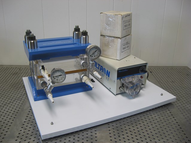 A tangential-flow filtration (TFF) cassette holder. Its clamped membrane cassettes concentrate the antibody and swap it into its final buffer — the UF/DF step that turns pure liquid into drug substance.
Tangential flow filtration system. Image by Kitmondo Marketplace, CC BY 2.0, via Wikimedia Commons.
A tangential-flow filtration (TFF) cassette holder. Its clamped membrane cassettes concentrate the antibody and swap it into its final buffer — the UF/DF step that turns pure liquid into drug substance.
Tangential flow filtration system. Image by Kitmondo Marketplace, CC BY 2.0, via Wikimedia Commons.
By now our antibody is genuinely pure. After capture, polishing, and two independent viral-safety steps (inactivation and filtration), almost nothing is left in the liquid but the protein we want. But that pure liquid still has two problems. There is too little antibody per drop — it is far too dilute to inject. And it is floating in the wrong liquid: a process buffer chosen to make filtration and chromatography work, not to keep the antibody happy for two years in a freezer. This final downstream step fixes both problems at once and hands us the drug substance (DS) — the concentrated, correctly-buffered protein that is the active heart of the medicine.
Think of making a pasta sauce. You start with a watery pot. You simmer it down so the flavor becomes rich and concentrated — that is the concentrating part. Then imagine you also need to swap the plain cooking water for a special seasoned broth that keeps it tasting perfect for months. You do both jobs in the same pot, without ever pouring the sauce out. That is exactly what this step does to the antibody: pack it tight, then bathe it in its final, protective liquid.
What this chapter covers
We will follow the pure but dilute antibody through its last downstream step, UF/DF, and watch a watery solution become a shelf-stable drug substance. You will see how a tangential flow filtration membrane concentrates the protein, how diafiltration washes out the old buffer and swaps in the final formulation, and the real numbers behind it — membrane cut-offs, the pressures and flux rates engineers actually run, how much fresh buffer a buffer exchange takes, and what goes into the formulation recipe. We will look closely at the one thing that can quietly ruin the batch here — aggregation — and at the compendial tests and GMP rules that keep it in check. Finally, we will see how a modern continuous line runs UF/DF inline instead of as a long batch pause.
What actually happens
The tool here is called UF/DF — ultrafiltration and diafiltration. Both are forms of tangential flow filtration (TFF), which uses a special membrane: a sheet riddled with pores so tiny that water and small molecules slip through, but the big antibody cannot.
The membrane: MWCO, TMP, and the concentration window
An IgG antibody weighs about 150 kilodaltons (kDa) — a kilodalton is just a unit for the mass of a molecule — and the membranes used here are rated to hold back anything above a chosen size, called the molecular weight cut-off (MWCO). For antibodies, that cut-off is typically 10–30 kDa, comfortably below the 150 kDa protein, so the antibody is reliably retained while water, salts, and small leftovers pass through [1]. The MWCO is a nominal rating, not a sharp wall: pore sizes scatter around it, which is why engineers pick a cut-off several-fold below the product mass — enough margin that monomer (one whole, intact antibody molecule) does not leak into the permeate, while smaller broken-off pieces — antibody fragments, or a stray light chain (one of the small sub-units that join together to make up the antibody) — being lighter, may still be partially lost.
Two streams flow out of any membrane:
- The retentate — what stays behind (our antibody). "Retained."
- The permeate — what passes through (water, salts, small leftovers). "Permeated."
Here is the clever part. If you pushed the liquid straight at the membrane, the antibody would pile up against it and clog the pores, like leaves jamming a storm drain. So instead the feed flows along the membrane surface, sideways, sweeping it clean, while a gentle pressure pushes water and small molecules through underneath. That sideways sweep is the "tangential" in tangential flow, and it is what lets a TFF system run for hours without choking. The control knob for how hard the pressure pushes is the transmembrane pressure (TMP) — the pressure difference across the membrane. Run it too low and almost nothing crosses; run it too high and you crush a dense protein layer against the surface (a problem called concentration polarization) that fouls the membrane and can damage the antibody. In practice operators hold TMP within a moderate window, often on the order of 0.5–2 bar (a bar is roughly the pressure of the atmosphere at sea level, so this is a gentle squeeze) for a typical antibody UF/DF, and tune both the TMP and the cross-flow rate together to push permeate through quickly while keeping the protein from clumping [2].
Ultrafiltration: concentrating the antibody and measuring flux
The first job is ultrafiltration (concentrate). Push water out as permeate; the antibody cannot leave, so it gets packed into less and less liquid. A feed that arrives at a few grams per liter can be concentrated many-fold; modern high-concentration formulations for subcutaneous injection (a small shot given just under the skin, where only about 1 mL of liquid fits) can reach 100 mg/mL or more, which is part of why aggregation control matters so much at this step.
The number that tells the operator how fast it is going is the permeate flux — the volume of liquid crossing the membrane per unit area per hour, written in L/m²/h. Flux is highest at the start, when the solution is thin, and falls steadily as the protein piles up and the cross-flow has to fight a thicker layer at the surface. Watching flux decline is how the team knows when the protein is concentrated and when the membrane is starting to foul; it is logged continuously alongside TMP and cross-flow velocity, and the cross-flow itself is often expressed as a velocity (on the order of hundreds of cm/h) that sets how vigorously the surface is swept.
Diafiltration: the buffer-exchange curve and diavolume math
In practice the two jobs are usually run in the order UF1 → DF → UF2: a first ultrafiltration to a moderate working concentration, then constant-volume diafiltration, then a final ultrafiltration up to the high target. The buffer exchange is deliberately done at the intermediate concentration, where the solution is still thin enough to give good flux — diafiltering at the final high concentration, where viscosity is punishing, would be painfully slow.
The second job is diafiltration (buffer exchange). Now add the final formulation buffer — the exact recipe of buffering salts and stabilizers the antibody will live in — at the same rate water is pulled off as permeate, so the volume stays steady. As fresh buffer flows in, the old process liquid washes out through the membrane. The amount of fresh buffer used is measured in diavolumes (one diavolume equals the volume of product in the system).
Buffer exchange follows a simple wash-out curve. Each diavolume removes a fixed fraction of whatever old buffer is left, so the curve is exponential: it falls fastest at the start and flattens as it approaches zero. Because the same fraction is removed each round, the amount left after N diavolumes follows the curve e^-N (here N is just the number of diavolumes, and e is the mathematical constant about 2.718 that always shows up when something decays by a steady fraction). One diavolume therefore leaves behind 1 divided by e — about 1/e (~37%) — of the old buffer, and the arithmetic compounds quickly:
| Diavolumes (N) | Old buffer remaining (e^-N) | Removed |
|---|---|---|
| 1 | ~37% | ~63% |
| 3 | ~5% | ~95% |
| 7 | ~0.1% | ~99.9% |
That last row is exactly the 99.9% removed figure. In practice teams commonly run on the order of 5–10 diavolumes to be sure the old buffer and residual process impurities are gone. That clean exponential assumes the species being washed out passes the membrane freely (a sieving coefficient near 1 — that coefficient is simply the fraction of a molecule that makes it through the membrane, where 1 means it passes completely and 0 means it is fully held back); anything the membrane partially retains washes out more slowly than the e^-N curve predicts. Round by round, the antibody ends up surrounded entirely by its final, protective liquid. The cleanest way to watch the exchange happen is to track conductivity — the solution's ability to carry electric current, which is dominated by dissolved salts. As the old salty process buffer washes out, conductivity decays along the same curve, which is exactly why an inline conductivity probe is the workhorse sensor for confirming a buffer exchange is complete.
 Diafiltration is an exponential wash-out: each diavolume of fresh buffer removes a fixed fraction of the old buffer, so conductivity decays steeply at first and about 7 diavolumes clears 99.9 percent.
Original diagram by the authors, created with AI assistance.
Diafiltration is an exponential wash-out: each diavolume of fresh buffer removes a fixed fraction of the old buffer, so conductivity decays steeply at first and about 7 diavolumes clears 99.9 percent.
Original diagram by the authors, created with AI assistance.
| UF/DF parameter | Typical value | Why it matters |
|---|---|---|
| Membrane cut-off (MWCO) | 10–30 kDa | Holds back the ~150 kDa antibody while water and salts pass |
| Transmembrane pressure (TMP) | 0.5–2 bar | Too low and little crosses; too high and the protein fouls the membrane and can clump |
| Final concentration | up to 100 mg/mL or more | Needed to fit a full subcutaneous dose into about 1 mL |
| Diafiltration | 5–10 diavolumes | About 7 diavolumes wash out ~99.9% of the old buffer |
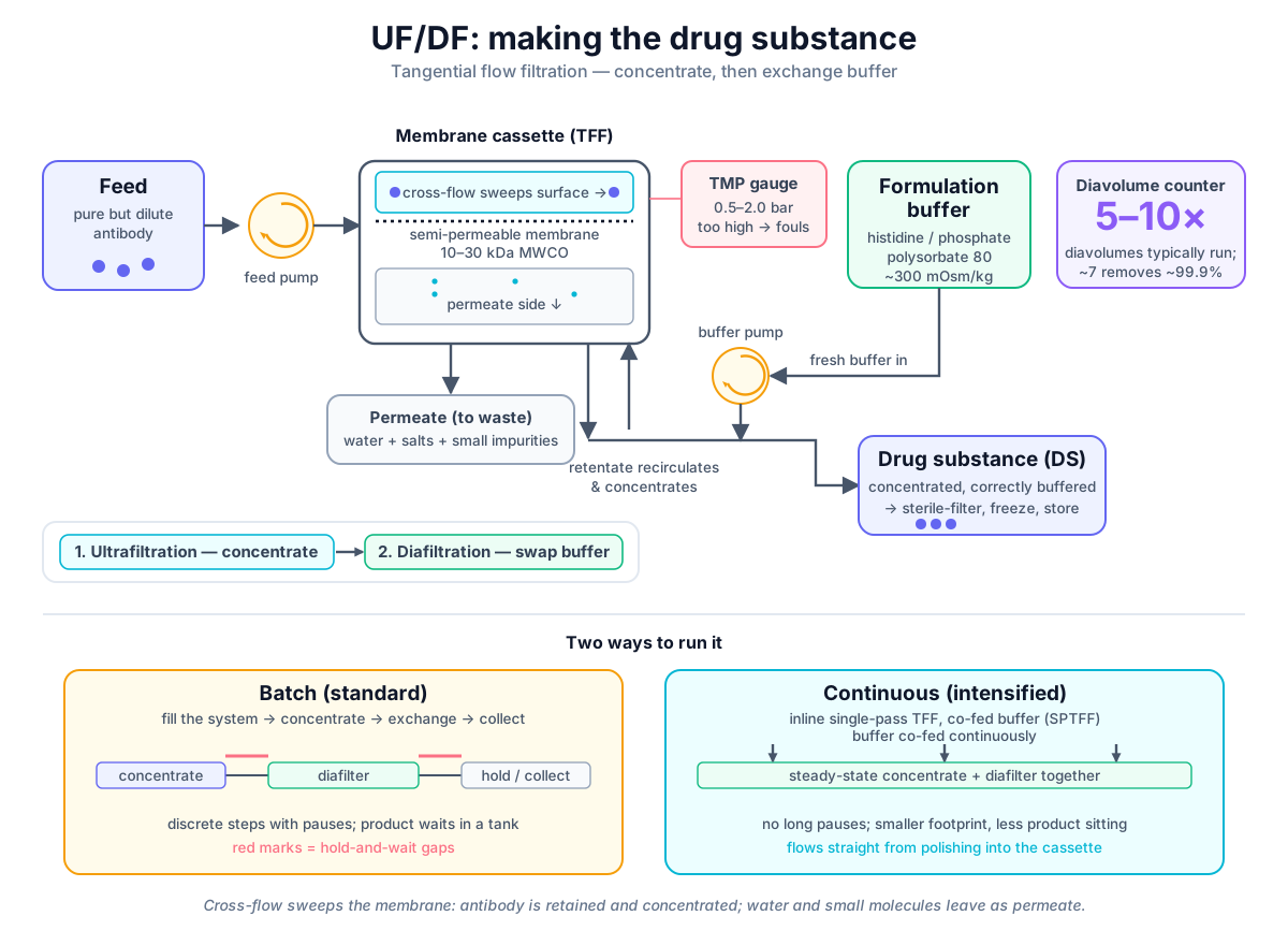 TFF system showing sideways cross-flow sweeping a permeable membrane, with retentate (concentrated antibody) exiting above and permeate (water + small molecules) below. Inset contrasts batch hold-and-exchange (hours-long pause) vs. continuous co-fed diafiltration (steady state with buffer pump).
Original diagram by the authors, created with AI assistance.
TFF system showing sideways cross-flow sweeping a permeable membrane, with retentate (concentrated antibody) exiting above and permeate (water + small molecules) below. Inset contrasts batch hold-and-exchange (hours-long pause) vs. continuous co-fed diafiltration (steady state with buffer pump).
Original diagram by the authors, created with AI assistance.

There is one last subtlety the diafiltration endpoint does not settle: the final concentration. Because some product is held up in the membrane and tubing, the system is deliberately overconcentrated slightly above target, then flushed and recovered with a buffer chase — which both rescues that held-up protein (step yields commonly run in the high-90s percent) and dilutes the pool. The pool is then adjusted back down to the exact target with formulation buffer in a final "spike" or QS step. This is why it is the final release assay, not the diafiltration endpoint, that fixes the drug substance concentration.
The result is concentrated, pure, correctly-buffered antibody. That is the drug substance. It is usually passed through one last sterilizing-grade filter, then filled into single-use bags or bottles and frozen for storage or shipping. This is the finish line of downstream processing.
Anatomy of one UF/DF cycle record
Before we move on to the formulation buffer, it is worth pausing on the data side of this step — a deliberate shift of lens that the companion books pick up in full. Every one of those parameters is logged, and together they form the data fingerprint of the step. A single TFF cycle is never recorded as a bare flux number — it is captured as a small identity card: a start timestamp; the TMP setpoint and the measured pressure; the cross-flow velocity in cm/h; the permeate flux in L/m²/h; a concentration proxy — an easy-to-measure signal that stands in for the real quantity, such as product density or refractive index, later confirmed by a direct lab assay (a test that measures the concentration itself); conductivity in mS/cm tracking how far the buffer exchange has gone; the temperature; and the hold time the concentrated protein has spent in the loop. Read together, these say not just what the value was but whether to trust it and which batch it belongs to.
 One UF/DF cycle record as an identity card: timestamped process parameters above, the deliverable concentration and buffer state in the green core, and the typed links that tie the cycle to its batch, phase, and skid.
Original diagram by the authors, created with AI assistance.
One UF/DF cycle record as an identity card: timestamped process parameters above, the deliverable concentration and buffer state in the green core, and the typed links that tie the cycle to its batch, phase, and skid.
Original diagram by the authors, created with AI assistance.
This is the moment a physical operation becomes a row of data. Each of those measured parameters is born at a sensor on the skid (the wheeled steel frame that carries the membrane, pumps, valves, and sensors as one self-contained TFF unit), and each reading becomes a timestamped, tagged data point in the batch's data shadow (the growing digital record of the physical lot that every measurement adds to). How that reading is captured, stamped with quality and time, and routed into the plant information systems is the subject of the data-management companion book — see where data is born, the data shadow, and where the plant systems pick up these readings. The structure of the identity card above — values bound to a batch, a phase, and a piece of equipment — is exactly the batch and equipment model that the open-source companion book implements in concrete schema and code, slotting into its reference architecture, and it is the downstream end of the same thread that begins when the upstream bioreactor first writes a titer reading. The violet panel on the card is not decoration: each line in it is a typed relationship a data model can read literally — this cycle runs-on this TFF skid, the cycle is-a diafiltration phase, the drug substance pool is derived-from this cycle. Naming those edges explicitly is what later lets a system trace a finished vial back to the exact membrane lot and cycle that made it; the ontology book builds that typed lineage in relations and genealogy and the class hierarchy behind it in classes and taxonomy. With the data side parked for the companion books, back to the liquid itself: what exactly is in that final buffer?
The formulation buffer: buffering agents, surfactants, tonicity
The formulation buffer is not plain salt water — it is a carefully chosen recipe whose only job is to keep a fragile protein intact for its whole shelf life. A few ingredients show up again and again [3]:
- A buffering agent to hold the pH steady, because antibodies are most stable in a narrow pH band (often around pH 5.5–6.5). Histidine (commonly 5–25 mM) and phosphate or acetate buffers are typical choices; a millimolar, mM, is just a measure of concentration.
- A surfactant such as polysorbate 80 or polysorbate 20, usually at a very low level (often around 0.01–0.05%), which coats the air-liquid and surface interfaces where antibodies otherwise unfold and stick. It guards against the mechanical stresses of pumping, filling, and shaking during shipping.
- A tonicity agent — frequently a sugar like sucrose or trehalose, or sodium chloride — adjusted so the solution's osmolality (its dissolved-particle concentration) sits near the body's own, roughly 300 mOsm/kg (milliosmoles per kilogram — a count of how many dissolved particles ride in each kilogram of water), so the injection is not painfully too concentrated or too dilute relative to blood.
One trap hides at high concentration: the final retentate is not a faithful copy of the diafiltration buffer. With 100+ mg/mL of antibody crowding the solution, the protein excludes volume and — because it carries charge — the Donnan effect shifts the small charged buffer species and the measured pH away from the buffer that was fed in. The mechanism is electrical balance: the big antibody molecules cannot cross the membrane but carry a net charge, so to keep each side electrically neutral they hold some of the small charged buffer ions close and push others away. That uneven split of the small ions is the Donnan effect, and the redistributed buffer ions nudge the measured pH a few tenths of a unit off the buffer that was fed in. Process teams account for this by deliberately offsetting the diafiltration buffer (for example, formulating to a slightly lower-than-target pH) so the finished high-concentration product lands on spec.
Get this recipe right and the antibody can survive months of freezing, thawing, and travel. Get it wrong and the protein slowly degrades — which brings us to why the whole step is treated so carefully. So far we have concentrated the antibody, washed it into its final protective buffer, and seen what one cycle records; that buffer recipe is what keeps the protein stable — which is exactly why the next risk to confront is aggregation.
Why it matters
The drug substance is the active medicine itself — everything before this step existed to produce it. If the final concentration is off, every later dose will be off. If the buffer exchange is incomplete, leftover process liquid can leave the antibody in a state where it is unstable.
The specific danger to watch is aggregation: individual antibody molecules sticking together into clumps. UF/DF is a high-stress moment for the protein — it is being squeezed to high concentration, pumped at speed, and pressed against a membrane — and all of that can coax some molecules to unfold and clump [2]. Aggregates are bad for two reasons. They are wasted medicine (a clumped antibody no longer works), and, more seriously, larger protein particles can be read by the patient's immune system as foreign, raising the risk of an unwanted immune response — what scientists call immunogenicity. The clinically worrying aggregates are not single misfolded molecules but larger assemblies, ranging from sub-micron clusters up through subvisible particles in the roughly 2–100 micrometer range and beyond.
Because of that, the antibody that comes out of UF/DF has to meet a defined set of specifications — the acceptance limits a drug substance must pass before it can move on. The international guideline ICH Q6B lays out the kinds of critical quality attributes a biologic must control, including purity, the fraction that is intact monomer versus aggregate, and residual impurities [4]. One of the routine release tests counts those clumps directly: USP general chapter <787>, Subvisible Particulate Matter in Therapeutic Protein Injections, sets the compendial method and limits for subvisible particles in injectable protein products [5] — a test that exists because this size range of particles was historically overlooked, a rationale we return to just below. It is the protein-specific companion to the older general injection test USP <788>: where <788> covers particulate matter in any injectable, <787> tailors the method to therapeutic proteins (smaller sample volumes, controls for the fact that protein aggregates are fragile and can be miscounted), so a protein drug substance is typically held to <787>. Done well, this step locks the antibody into a stable state and passes those tests cleanly; done badly, it shows up as aggregates and rejected batches.
How high-concentration stress fails the batch
It is worth being concrete about exactly how this step throws away a batch, because the failure is quiet. The danger window is the end of ultrafiltration, where the protein is at its most crowded and most stressed. Squeezed to 100 mg/mL or more, pumped repeatedly through the recirculation loop, and pressed against the membrane, a small fraction of molecules unfold at the interfaces and seed (nucleate) new aggregates — one unfolded, sticky molecule acts as a starting point that others clump onto. These do not announce themselves — the solution can still look clear by eye — but they populate the subvisible particle range, the protein particles larger than about 2 micrometers that the human eye cannot resolve.
Crowding brings a second, blunter limit: above roughly 100–150 mg/mL the solution's viscosity climbs steeply, and it — not aggregation alone — often sets the highest concentration and flux the process can actually reach. That viscosity wall is why high-concentration formulations lean on charge-shielding excipients (added, inactive helper ingredients in the formula) such as arginine: when packed this tightly the antibodies repel one another through their surface charges, and that mutual shoving is part of what thickens the solution; arginine screens those charge repulsions so the molecules slide past each other more easily and the liquid flows better. Open-channel cassettes that resist clogging by a thick feed help too. A foundational commentary by Carpenter and colleagues argued that this size range had been historically overlooked and that such particles deserve direct attention precisely because of their potential to provoke an immune response [9].
When a batch crosses the USP <787> limit on subvisible counts, it fails release and is rejected — there is no rework that un-aggregates a protein. Regulators treat particulate control as a serious matter: the FDA's own guidance on inspecting injectable products makes clear that visible and subvisible particulate contamination is grounds for rejecting a lot, and particulate-related deficiencies are a recurring theme in agency findings on parenteral manufacturing [10]. The practical consequence is that the seemingly mundane parameters on the cycle record — hold time, temperature, how hard the protein was pumped — are not paperwork. They are the levers that decide whether the most expensive material in the building passes or is poured down the drain.
It is also worth knowing what comes next. The drug substance is not the final medicine a patient receives. It still has to be turned into the drug product (DP) — the actual vial or prefilled syringe — during formulation and fill-finish. DS is the purified active ingredient at its correct concentration and buffer; DP is the packaged, labeled, ready-to-use dose. The line between them is exactly the line this chapter ends on.
In the real world
Behind the friendly pasta-sauce analogy sits a wall of named equipment, validated procedures, and compendial standards.
Single-use vs reusable systems and validated parameters
Commercial UF/DF runs on TFF skids — pump-and-membrane systems built around either flat-sheet cassettes or hollow-fiber modules. Major suppliers include Cytiva (Kvick and UFP cassettes), Sartorius (whose Hydrosart stabilized-cellulose cassettes are widely used), Pall (now part of Danaher, maker of hollow-fiber and Cadence single-pass modules), and Repligen (whose KrosFlo hollow-fiber systems are a downstream staple). The membranes are commonly polyethersulfone (PES) or regenerated cellulose, chosen for low protein binding and chemical robustness, and selected at a 10–30 kDa MWCO to retain the antibody.
The size of the unit is set by membrane area — how many square metres of membrane the skid carries — which in turn is sized from a loading target: how many grams of antibody each square metre of membrane can process in one batch (the g/m² load). Process development runs a small cassette to find a load and a flux the membrane can sustain without fouling, then scales up by holding that load constant — a clinical batch might run a few square metres while a commercial batch runs tens, with the same g/m². That single number is one of the parameters frozen at technology transfer and re-checked during process qualification (the formal IQ/OQ/PQ exercise that proves the installed equipment is installed correctly, operates as intended, and performs consistently at full scale). On a reusable skid the membranes are not discarded after one batch but regenerated — cleaned and stored in a preservative between uses — and each cassette is rated for a validated number of re-use cycles, after which it is replaced.
The choice between reusable stainless skids and single-use flow paths shapes both the cleaning burden and the validation work. A reusable system must be cleaned, sanitized, and re-qualified between batches, and its cleaning has to be proven not to carry material from one product to the next. Increasingly the cassettes and flow paths are single-use, so there is no large stainless system to clean and re-validate between batches — the path is installed, used once, and discarded.
Either way, the UF/DF process is validated and run to fixed limits. Every filter in the fluid path is governed by cGMP — current Good Manufacturing Practice, the legally binding rules that require every batch to be made the same controlled, documented, traceable way. FDA 21 CFR 211.72 specifically governs the filters used in processing injectable products, including the requirement that the final sterilizing filter be a non-fiber-releasing 0.2-micrometer filter [6] — pores that small physically strain out bacteria so the stream comes through sterile, and "non-fiber-releasing" means the filter must not shed its own fibers into the drug — the same compendial filter logic that governs the upstream viral-safety filtration step. The membrane's integrity is verified both before and after use by an air-based diffusion (forward-flow) or pressure-hold test measured against a validated specification: the operator wets the membrane, pushes air against it at a set pressure, and measures the air flow or pressure decay — an intact membrane holds water in its tiny pores and lets only a small, predictable trickle of air through, so an oversized hole shows up as too much air flow. A failed post-use integrity test quarantines the batch pending investigation. Parameters like TMP, cross-flow rate, temperature, protein concentration, and hold time are documented and held inside qualified ranges in the batch record — the controlled, signed document that records every step and measurement for one batch and is the legal proof of how it was made. Those entries carry a data-integrity expectation with a name: each must be ALCOA+ — Attributable, Legible, Contemporaneous, Original, and Accurate, plus complete, consistent, enduring, and available — and the software on the skid that captures the cycle is itself validated and access-controlled (electronic-signature and audit-trail rules, often cited as 21 CFR Part 11), with the field shifting from exhaustive scripted computer system validation (CSV) toward risk-based computer software assurance (CSA). The sister data book treats these in full in data integrity and ALCOA+ and the move from CSV to CSA. Hold time and temperature matter because a concentrated protein left warm for too long is a protein that starts to aggregate.
Modern continuous UF/DF and inline PAT
In the standard commercial process, UF/DF is a single batch at the very end of the downstream line: fill the system, concentrate, exchange buffer over its diavolumes, recover, and freeze — a process that can pause the line for hours while the product sits in the recirculation loop. Fed-batch upstream (an upstream strategy where nutrients are fed into the culture in stages, covered in the earlier chapters) feeding into batch downstream still makes the great majority of approved monoclonal antibodies (mAbs) today. But the emerging direction is continuous and intensified processing, in which the product never sits still in big tanks and instead flows steadily from one operation to the next [7]. In that mode, single-pass TFF (SPTFF) lets the antibody concentrate in one pass through a long membrane path rather than recirculating for hours, and an inline diafiltration stage can be fed continuously. Wired together, this lets the antibody flow more or less directly from the final polishing column through inline concentration and buffer exchange without an intermediate hold tank — which shortens the time the protein spends at high concentration and shrinks the equipment footprint.
Process analytical technology (PAT) sensors — inline gauges for conductivity (a proxy for buffer exchange) and ultraviolet absorbance or turbidity (a proxy for protein concentration — proteins absorb ultraviolet light and scatter visible light in proportion to how much protein is present, so a stronger signal means more antibody) — watch the streams in real time and let the system adjust feed and buffer rates to hold the target steady. Those same continuous PAT streams are what feed the statistical process control and continued process verification machinery that proves, batch after batch, that the step stays inside its qualified window. They are also where this step starts to add a predictive layer: because the true concentration is slow to measure in the lab but the inline UV and conductivity signals track it closely, a soft sensor (a model that infers a hard-to-measure quantity from easier signals — here estimating concentration or the diafiltration endpoint in real time) can call the endpoint before the lab assay confirms it and tune feed and buffer rates on the fly. The ML companion book works exactly this step in UF/DF and drug substance soft-sensing. A foundational white paper on continuous bioprocessing makes the case that this kind of integration brings smaller equipment, lower cycle times, and a more streamlined plant [8].
Key terms
- Drug substance (DS) — the pure, concentrated, correctly-buffered antibody; the active medicine before packaging.
- UF/DF — ultrafiltration plus diafiltration; the step that concentrates the antibody and swaps its liquid.
- Tangential flow filtration (TFF) — filtering where liquid flows sideways along the membrane to keep it from clogging.
- Membrane — a sheet with pores small enough to hold back the antibody but let water and small molecules through.
- Molecular weight cut-off (MWCO) — the size limit a membrane retains; for antibodies, typically 10–30 kDa, well below the ~150 kDa IgG.
- Retentate — the liquid kept behind the membrane (the antibody).
- Permeate — the liquid that passes through the membrane (water and small molecules).
- Permeate flux — the volume of liquid crossing the membrane per unit area per hour (L/m²/h); it falls as the protein concentrates and signals fouling.
- Cross-flow velocity — how fast the feed sweeps along the membrane surface (often hundreds of cm/h), keeping the membrane from clogging.
- Conductivity — the solution's ability to carry current, dominated by dissolved salts; tracked inline as the live proxy for how far the buffer exchange has progressed.
- Diafiltration — washing out the old liquid and replacing it with the final formulation buffer at constant volume.
- Diavolume — a measure of how much fresh buffer is used in diafiltration; one diavolume equals the product volume in the system.
- Formulation buffer — the final, stabilizing liquid the antibody is placed into (buffering agent, surfactant, tonicity agent).
- TMP (transmembrane pressure) — the pressure difference across the membrane that controls how fast liquid crosses it.
- Osmolality — the concentration of dissolved particles in the solution, set near the body's ~300 mOsm/kg for injection.
- Aggregation — antibody molecules sticking together into clumps; a key risk during high-stress concentration.
- Immunogenicity — the chance that aggregates or particles trigger an unwanted immune response in the patient.
- Subvisible particles — protein particles too small to see by eye, counted and limited by compendial tests such as USP <787>.
- Membrane area / loading — the square metres of membrane on the skid, sized from a grams-per-square-metre (g/m²) load that is held constant on scale-up.
- Single-pass TFF (SPTFF) — concentrating product in one continuous pass through the membrane, used in intensified processing.
- Process analytical technology (PAT) — inline sensors that measure the process in real time so it can be adjusted on the fly.
- Soft sensor — a model that infers a hard-to-measure quantity (here, concentration or the diafiltration endpoint) from easier inline signals like UV and conductivity.
- ALCOA+ — the data-integrity expectation that every record be Attributable, Legible, Contemporaneous, Original, and Accurate (plus complete, consistent, enduring, available).
- Drug product (DP) — the finished, packaged medicine (vial or syringe) made from the drug substance.
Where this leads
The drug substance is now bottled or bagged and frozen — pure, concentrated, and resting in its protective buffer. But it is still bulk material, not yet a dose a nurse can draw up. In the next chapter, formulation and fill-finish, we follow the drug substance into the sterile filling line, where it is finalized, filtered, and dispensed into the actual vials and syringes that become the drug product — the medicine that finally reaches the patient.