Viral safety step 2: filtering viruses out
📍 Where we are: Stop 16 of 21 — the second viral-safety step. We have weakened viruses with acid; now we physically strain the liquid through a filter so fine that even a virus cannot slip through.
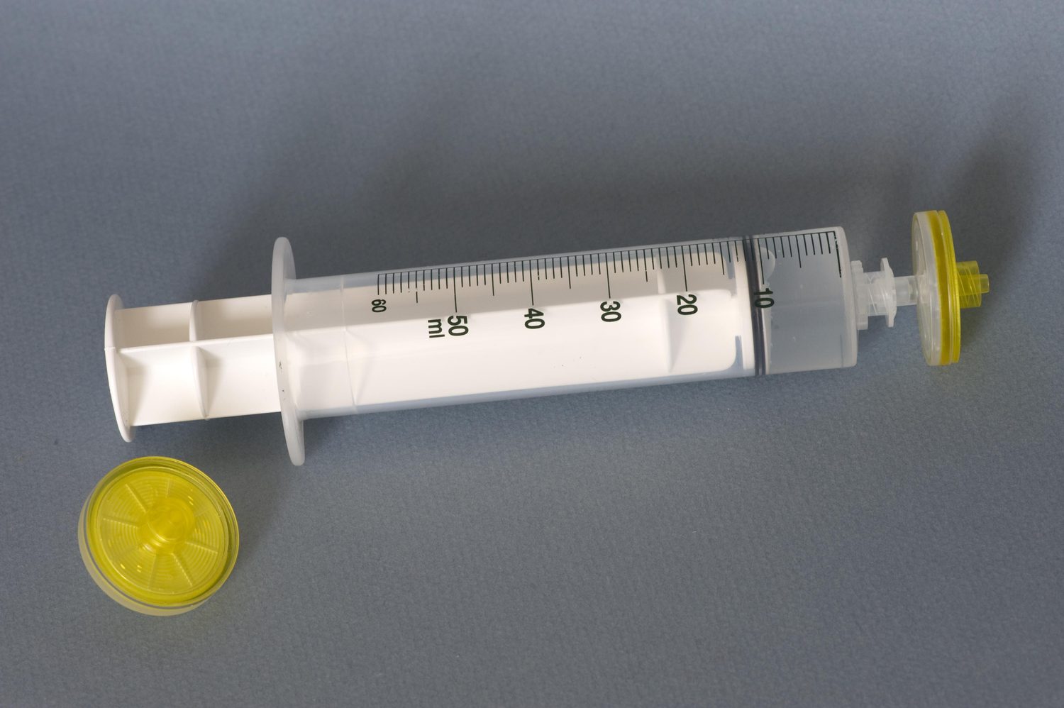 A membrane filter capsule. Virus filtration works on the same principle at production scale: fluid is pushed through a membrane whose pores are small enough to let the antibody pass but trap anything virus-sized.
Membrane filter capsule. Image by Stephane Lesbats (IFREMER), CC BY 4.0, via Wikimedia Commons.
A membrane filter capsule. Virus filtration works on the same principle at production scale: fluid is pushed through a membrane whose pores are small enough to let the antibody pass but trap anything virus-sized.
Membrane filter capsule. Image by Stephane Lesbats (IFREMER), CC BY 4.0, via Wikimedia Commons.
We have already weakened viruses with acid in the low-pH step. Now we do something completely different: we physically remove any remaining viruses by trapping them in a filter with pores almost too small to imagine. The antibody molecule is small enough to pass; most viruses are too big, so they get stuck. Two unlike steps, two different ways to fail — and that is exactly the point.
Imagine a sieve so impossibly fine that water and tiny grains of sugar flow right through, but even a speck of dust gets caught. Our antibody is the sugar. A virus is the speck of dust. We push the liquid through the sieve, and only the clean, virus-free liquid comes out the other side. Real life adds one twist: a few specks are nearly the same size as the sugar, so the sieve has to be designed and proven very carefully to catch them too.
What this chapter covers
This chapter explains the second of two viral-safety steps: virus-retentive filtration (often called nanofiltration). We will walk through how a filter with pores around 20 nanometers wide lets the antibody through while holding viruses back, look at the real machines and membranes used in industry, and see the actual numbers operators watch — pressure, flow, and the slow clogging called fouling. We will see how companies prove the filter works by deliberately spiking it with viruses, how every batch is checked afterward, what a single filter costs, and how this once-paused step is being reshaped into a continuously flowing stage in modern facilities. Along the way we will fix a common oversimplification: viruses are not held back by size alone.
How the nanofilter works
The virus-retentive nanofilter: physics and design
The key tool here is a virus-retentive nanofilter — a special filter whose holes (pores) are around 20 nanometers wide. A nanometer is one-millionth of a millimeter, so these pores are fantastically small. The main trick is a difference in size: a single antibody molecule is only about 10 nanometers across, so it slides through easily, while most viruses are larger and cannot fit.
This is called size exclusion — separating things mainly by how big they are, like a security checkpoint that turns away anything above a strict size limit. But here is the honest version that the simple sieve picture leaves out: real filters do not catch viruses by size alone. Three things work together. First, steric hindrance — the absolute size cutoff, around 15 to 20 nanometers, that simply blocks anything bigger. Second, in some membranes, a mild electrostatic interaction: viral shells tend to carry a negative surface charge (their protein coat, like the antibody and impurity proteins sorted by charge in the polishing step, is net-negative at the near-neutral pH used here), and certain membrane regions carry a positive one, so the virus is gently pulled toward and held by the membrane. Third, a small amount of adsorption — viruses sticking to the membrane surface as they pass. Together these give the filter more grip than pore size would suggest, which matters most for the smallest, hardest-to-catch viruses [2].
Here is the sequence:
- The antibody liquid — already through Protein A capture and the polishing step — is gently pushed toward the nanofilter under steady pressure.
- The liquid is forced through the tiny pores. The push is measured as transmembrane pressure (TMP) — the pressure difference across the membrane — and is kept in a careful window — illustratively 0.5 to 2.5 bar (a bar is roughly one atmosphere, the pressure of the air around us, so 2.5 bar is about two and a half times that; nanofilters frequently run around 1.0 to 1.5 bar), though the exact window is vendor- and product-specific and fixed during validation. Some hollow-fiber filters (many fine tubes the liquid flows through) run well under 1 bar; flat-sheet filters (stacked membrane sheets) can run higher. Push too hard and you risk damaging the membrane or driving something through that should have stayed behind.
- Antibody molecules pass through and are collected on the far side as clean, filtered product. A run typically lasts a few hours — often 2 to 6 — depending on how much liquid there is and how concentrated the antibody is.
- Any viruses too large to fit stay behind, trapped on the membrane, and are discarded with the spent filter.
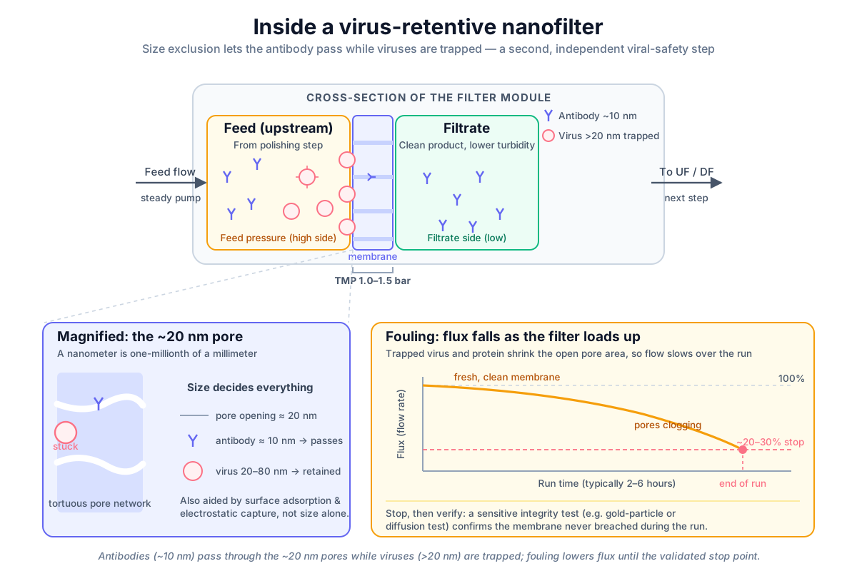 Virus-retentive nanofilter architecture and mechanism: antibodies (10 nm) pass through, most viruses (larger than the pores) are trapped — and even borderline 18 to 26 nm parvoviruses are held by added electrostatic and adsorptive grip — while membrane fouling reduces flow rate over time.
Original diagram by the authors, created with AI assistance.
Virus-retentive nanofilter architecture and mechanism: antibodies (10 nm) pass through, most viruses (larger than the pores) are trapped — and even borderline 18 to 26 nm parvoviruses are held by added electrostatic and adsorptive grip — while membrane fouling reduces flow rate over time.
Original diagram by the authors, created with AI assistance.

Because the filter works mostly by a physical barrier rather than by chemistry, it does not care whether a virus is fragile or tough — if it is too big, it cannot get through. That is exactly why this step is so valuable. (One small caveat for the curious: the process conditions — pH, dilution, osmotic stress in the feed — can on their own knock out some delicate viruses before they ever reach the membrane, so the filter occasionally gets a little unearned help. The validated retention claim, though, rests on the membrane doing the work, not on that lucky bonus.)
Anatomy of a 20-nanometer membrane
It helps to stop thinking of the membrane as a flat screen with round holes and instead see it as a designed object with several properties working at once. The pore-size rating sets the absolute cutoff; the polymer (the tough plastic-like material the membrane is made of) — usually polyethersulfone (PES) or polyvinylidene fluoride (PVDF) — gives the membrane its chemical toughness and its very uniform fine pores; the surface charge adds an electrostatic grip on top of pure geometry; and the membrane-area rating (in square meters) sets how much liquid the module can take before it fouls. The card below lays out the same membrane as an identity record: each attribute, its value, and what it buys you.
 The membrane as an identity card: a 20-nanometer cutoff in a PES or PVDF polymer, mildly positive at its surface, retaining 3 to 4 logs of virus by three mechanisms at once — steric, electrostatic, and adsorptive.
Original diagram by the authors, created with AI assistance.
The membrane as an identity card: a 20-nanometer cutoff in a PES or PVDF polymer, mildly positive at its surface, retaining 3 to 4 logs of virus by three mechanisms at once — steric, electrostatic, and adsorptive.
Original diagram by the authors, created with AI assistance.
Measuring and proving retention
Log reduction value: measuring retention
How do we put a number on "this filter removes viruses"? Engineers use the log reduction value (LRV) — a measure of how many viruses go in versus how many come out, counted in factors of ten. An LRV of 3 means a thousand-fold reduction (for every 1,000 viruses in, 1 comes out). An LRV of 4 means ten-thousand-fold. A virus-retentive filter typically delivers roughly 3 to 4 LRV — a 1,000- to 10,000-fold reduction — and a well-designed one can do considerably more.
A single step usually does not have to carry the whole burden. Regulators expect the overall process — low-pH inactivation, chromatography, and filtration combined — to deliver a large cumulative clearance, and individual robust steps are commonly credited with 4 or more logs each. Because the logs add up across orthogonal steps, a process that earns 4 logs at the acid step and another 4 at the filter has demonstrated an 8-log (hundred-million-fold) clearance overall — the kind of headroom regulators look for. The expectations and the way to demonstrate them are laid out in international guidance and in pharmacopeial chapters that the whole industry follows [1] [3]. The arithmetic is only as good as its weakest credited step, though, which is why the filter's claim has to be earned, not borrowed from a sister product.
But there is a catch, and it is the most important honest point in this chapter. Not all viruses are the same size. The biggest worry is the family of small, non-enveloped viruses — hard protein shells with no fatty coat — and the smallest of these, parvoviruses, are only about 18 to 26 nanometers across. That puts them right at the edge of a 20-nanometer filter's cutoff. Put the three numbers in one breath: a 20-nanometer pore sits comfortably above the 10-nanometer antibody but right on top of the 18-to-26-nanometer parvovirus — which is exactly why parvovirus, not some larger virus, is the design challenge. They are not comfortably blocked by size the way a large virus is; they sit on the borderline. This is precisely why the extra electrostatic and adsorptive grip matters, and why a manufacturer can never simply assume a filter stops parvovirus. They have to prove it, virus by virus.
Proving it works: spiking studies with model viruses
There is a reason this matters specifically for our running mAb (monoclonal antibody — the antibody drug we are making). Our antibody is made by CHO cells — Chinese Hamster Ovary cells, the workhorse production cell line of the industry — and CHO cells endogenously express retrovirus-like particles (RVLPs) — non-infectious, roughly 80-to-100-nanometer particles budded from the genome — so under ICH Q5A (the international viral-safety guideline every biologic process is built to satisfy) every CHO mAb process must demonstrate retrovirus clearance even when no adventitious agent is ever found. That is why a retrovirus model always appears in the panel alongside the small-parvovirus worst case.
You cannot release a medicine on the promise that a filter "should" work. So before a process is approved, manufacturers run viral clearance validation, also called spiking studies. The idea is blunt: deliberately add a large, known quantity of real virus to a sample of the process stream, run it through the filter under the worst-case conditions the real process might ever see, and measure how much virus survives. That measured reduction becomes the claimed LRV. "Worst-case" is doing real work in that sentence — the study is run at the highest pressure, the most fouling-prone feed, and the most extreme processing time the commercial process is allowed to reach, precisely so that the claim still holds on the worst day a real batch can have. This work is done at small (scaled-down) scale by specialist labs, on a model of the production filter that has been shown to behave like the full-size one, because spiking a full manufacturing batch with live virus is neither safe nor sensible.
To do this fairly, scientists challenge the filter with carefully chosen model viruses that stand in for the things that might realistically contaminate a cell line. A good panel must probe both ends of the size range — an easy large virus and the hard small-parvovirus borderline — using real model viruses plus well-characterized bacteriophages (viruses that infect bacteria and are harmless to people) as safe, standardized stand-ins, since handling live virus at scale is hazardous and expensive [4]. One small-virus model is not an arbitrary choice: MVM stands for minute virus of mice, and CHO is a Chinese hamster line — a hamster is a rodent, like a mouse — so a well-characterized mouse parvovirus (MVM, ~18 to 24 nm) is the conventional small-virus model regulators expect for a rodent-derived CHO process, making it the design-driving challenge agent. The table below names each model, its size, and the role it plays.
| Model virus | Approx. size | Type | Role in the challenge |
|---|---|---|---|
| Porcine parvovirus (PPV) | 18–26 nm | Non-enveloped | Probes the hardest small-virus case |
| Minute virus of mice (MVM) | ~18–24 nm | Non-enveloped | The standard small-virus model for CHO processes (endogenous parvovirus risk) |
| Pseudorabies virus (PRV) | Large | Enveloped | Stands in for the easy, large end |
| PR772 | ~80 nm | Phage (safe surrogate) | Tests large-virus retention |
| PP7 | ~25 nm | Non-enveloped phage (safe surrogate) | Probes the small-parvovirus borderline |
| PhiX174 | ~27 nm | Non-enveloped phage (safe surrogate) | Probes the small-parvovirus borderline |
Bacteriophages are listed by their safe-surrogate role; they are viruses that infect bacteria and are harmless to people.
The experimental design — how many runs, what controls, how to calculate the reduction, how to handle the worst case — is spelled out in detailed regulatory and pharmacopeial guidance so that one company's "4 logs" means the same thing as another's [1] [3].
Running the filter and proving the membrane held
Running the filter: transmembrane pressure and flux decline
A virus filter is not a passive screen you set and forget. As the run proceeds, viruses, protein, and other particles pile up on the membrane and partly plug its pores. The effective open area shrinks, and the flow rate through the filter — its flux — slowly falls. This gradual clogging is called membrane fouling, and the matching drop in throughput is flux decline.
Operators watch flux closely, because fouling is not just an efficiency problem — it can change how the filter retains viruses. A filter pushed far past its safe range, or run at the wrong pressure, may behave unpredictably. So the process is stopped well before flux falls too low — commonly when it has declined to roughly 20 to 30 percent of where it started, or when a preset volume has been processed, whichever comes first. The pressure and flow envelope and the maximum hold are defined in advance and recorded for every batch, never improvised on the day [2].
Notice what is happening underneath the operator's eye: TMP and flux are not just gauges to glance at, they are signals sampled every few seconds, trended, and stored. That live trend is the physical birth of a data point — one more tagged measurement joining this batch's data shadow (the complete digital record that grows alongside the physical product). The same pressure-and-flux story is told three ways across this trilogy — as a physical run here; as a born data point with quality and lineage (a documented trail of where the value came from) in the companion Data Management book's plant-information-systems chapter — see also where process data is born and the biologic's data shadow; and as a concrete database row a query can pull back in the Open-Source Systems chapter on downstream chromatography and filtration. When that stored value is later tied back to this filter, this batch, this step, it forms a typed relationship — a derived-from / runs-on link of the kind the Ontology book treats as a genealogy edge, so the number is never an orphan. The diagram below follows one run along exactly that chain — sensor to historian to verdict.

Post-use integrity testing: pressure decay, gold particle, diffusion
Integrity tests answer one question: did the membrane stay intact? When the run is finished, the work is still not done — you have to confirm the membrane never developed a flaw during use. This is post-use integrity testing. (Some processes also run a pre-use, post-sterilization integrity test, or PUPSIT, before the batch ever touches the filter. Whether such a pre-use test usefully catches a defective filter, or whether the wetting liquid needed to run it can temporarily plug a small flaw and hide it, is an ongoing regulatory debate sharpened by the EU's Annex 1 (the European GMP rules for sterile medicines). Most mAb processes rely on the post-use test described here.)
The easiest way to picture the principle is the classic bubble-point test: the membrane is wetted, gas pressure is slowly raised on one side, and the pressure at which gas first pushes through reveals the size of the largest pore. A membrane with an intact, fine pore structure holds out to a high, expected pressure; one with a defect lets gas through too early and fails. That classic bubble-point method (standardized for microfiltration membranes — those with much larger, micron-scale pores, roughly a thousand times wider than a virus pore — as ASTM F316) works on micron-scale pores, but a true 20-nanometer virus pore would need impractically high pressures to bubble through.
So virus filters are instead confirmed with more sensitive, vendor-specified tests tuned to the nanometer scale — for example a gold-particle test, a binary-gas test, or a diffusion / forward-flow or pressure-decay test — each chosen to flag a flaw far smaller than a classic bubble point could. These are not arbitrary checks: the vendor has correlated a specific test result (a maximum allowed gas flow, a maximum pressure decay over a set time) against the actual virus retention measured in the spiking studies, so that a passing number is a stand-in for "this membrane still delivers its claimed LRV." Concretely, the acceptance logic is a simple inequality: if the vendor sets the limit at, say, a maximum diffusive gas flow correlated to the claimed LRV, a measured flow below that limit passes and a flow above it fails — one number, read against one threshold. That is what lets a quick physical test at the end of a run substitute for re-running a virus challenge on every batch.
The filtered product is also checked for cleanliness — turbidity and subvisible particle counts (tiny particles too small to see) against pharmacopeial limits — to confirm nothing unexpected came through. Only a batch whose filter passed integrity and whose product passed these checks moves forward. Every one of these results — the recorded TMP and flux trend, the integrity verdict, the particle counts — becomes part of the permanent batch record, and in a computerized plant it must be captured under the same electronic-records rules that govern any GMP (Good Manufacturing Practice — the legally enforced rules for how medicines are made) data. Each such record is held to the ALCOA+ principles — that the data be Attributable, Legible, Contemporaneous, Original, and Accurate (plus complete, consistent, enduring, and available) — the shorthand regulators use for what makes a record trustworthy. The Data Management chapter on data integrity and ALCOA+ and the chapter on Part 11 and Annex 11 walk through these rules in full, and the Open-Source Systems chapter on contextualization shows how a raw sensor stream is tied back to this batch, this filter lot, this step so the verdict means something.
When fouling goes wrong: the plugged-membrane failure mode
The honest way to understand a control limit is to watch it being breached. Picture a batch whose feed carried an unusually high load of antibody aggregate — clumped, sticky protein that the upstream steps did not fully trim. The membrane pre-loads far faster than the validated model predicts. Flux collapses early, and to keep the liquid moving an operator (or an automated loop) lets transmembrane pressure climb. It crosses the validated pressure ceiling — here taken as 2.5 bar, the top of this product's qualified envelope, not a universal industry constant. Now two things are wrong at once: the run is outside its proven envelope, and a membrane stressed above its qualified pressure can no longer be assumed to retain viruses the way the spiking studies showed.
This is a hard stop, not a judgment call. The batch goes on hold, an investigation opens, and the post-use integrity test is read with extra scrutiny because the very excursion that plugged the filter could also have strained it into a breach. The discipline here is not optional housekeeping — it is the core logic of aseptic and sterile-by-filtration processing that the US FDA lays out in its aseptic-processing guidance, and it sits on top of binding US federal law that a licensed biologic meet its purity standard on every batch (21 CFR Part 610) and the international viral-safety guideline that virus clearance be validated and held within proven limits (ICH Q5A(R2)) [8] [6] [1]. A filter that ran outside its envelope did not "probably still work"; it has to be proven, or the batch does not ship.
Orthogonal defense in depth and commercial membranes
Why it matters: orthogonal defense in depth
The low-pH step is great at destroying enveloped viruses — viruses wrapped in a fatty outer coat that acid can break apart. But non-enveloped viruses have a hard protein shell and no fatty coat, so acid barely touches them. These tough, often very small viruses could slip past the first step.
Viral filtration is the safety net aimed at them. It does not rely on chemistry, so it works on the very viruses the acid step might miss — provided, as we saw, the filter has been proven to catch even the borderline-sized ones.
This is the heart of an idea called defense in depth: using two independent steps that work on totally different principles. Recall that the low-pH step destroyed enveloped viruses by chemistry; this step removes the survivors mainly by size. For a virus to survive the whole process, it would have to defeat both — and a virus that shrugs off acid is still confronted by the membrane, while a virus small enough to test the membrane's limit is usually fragile enough for acid to destroy. Stacking two unlike, orthogonal steps gives extremely high assurance that the final medicine carries no infectious virus. For a drug injected into a sick patient, that assurance is not a nice-to-have; it is the law and the ethics of the work [1].
In the real world: commercial membranes and the continuous shift
Virus filtration is a mature, well-supplied corner of bioprocessing, and several vendors build the membranes and systems used worldwide. Common names a newcomer will hear include Asahi Kasei's Planova family (the original hollow-fiber virus filters), Millipore (Merck) Viresolve, Sartorius Virosart, Pall virus-removal filters, and Repligen Pegasus modules. Two facts about each are worth keeping straight — its structural format (the physical shape of the membrane) and its polymer (the plastic it is made of) — so the table below lays out, for each product family, the vendor, format, and polymer in one place rather than scattering them across the prose.
| Product family | Vendor | Format | Polymer |
|---|---|---|---|
| Planova | Asahi Kasei | Hollow-fiber | Regenerated cellulose (BioEX grade: PVDF) |
| Viresolve | Millipore (Merck) | Flat-sheet / cassette | PES |
| Virosart | Sartorius | Flat-sheet / cassette | PES |
| Pall virus-removal | Pall | (vendor-specific) | (vendor-specific) |
| Pegasus | Repligen | (vendor-specific) | (vendor-specific) |
Format and polymer are listed only where the page names them; "vendor-specific" marks a value that varies by product grade and is not fixed in this overview.
In narrative terms, the families split into two structurally different formats. Hollow-fiber filters like the classic Planova line feed liquid through the lumen of many fine fibers (inside-out or outside-in), while flat-sheet / cassette products like Viresolve and Virosart stack pleated or layered membrane sheets — and the geometry shapes how flux distributes across the membrane and how throughput is rated. The polymers are all rugged, uniform-pore plastics — broader than the two most common modern choices named earlier — such as regenerated cellulose (the historically dominant virus-filter chemistry, used in the classic Planova grades), polyethersulfone (PES), or polyvinylidene fluoride (PVDF) (used in flat-sheet products and the Planova BioEX grade) — materials chosen because they are chemically tough, low-fouling, and can be made with extremely uniform fine pores.
Filters are sized by membrane area, and operators run them within a flux envelope. Flux is conventionally expressed in LMH — liters per square meter of membrane per hour — and a typical virus-filter operating flux is roughly 30 to 50 LMH, scaled to the batch — a brisk, push-it-through pace. (Continuous and intensified runs, which trade speed for gentleness, can sit much lower, around 7 to 10 LMH — a deliberately slow trickle.)
These are precision components, and they are not cheap. A single virus-filtration module can cost on the order of 15,000 to 40,000 US dollars depending on its membrane area and grade. That price shapes real decisions. Some systems are strictly single-use — installed, run once, then discarded with the trapped virus — which is simple and removes any worry about carryover but adds consumable cost to every batch. Others can be reused after a validated cleaning and sanitization cycle (for example a hot caustic wash followed by storage in dilute ethanol), trading a more complex cleaning-validation burden for lower per-batch cost. Neither is automatically "better"; it is a genuine economics-versus-simplicity trade-off that each manufacturer weighs.
All of this sits inside a firm regulatory frame. In the United States, federal law (21 CFR Part 610) requires that licensed biologics meet standards for safety, purity, and potency — which legally obligates a manufacturer to validate and then monitor viral-clearance steps like filtration for every batch [6]. In Europe, the European Pharmacopoeia sets a parallel expectation that validated virus-removal and inactivation procedures be applied and that viral-safety risk be assessed for any product made with cells of human or animal origin [5]. This is current Good Manufacturing Practice (cGMP) — the up-to-date, legally enforced rulebook for how medicines must be made — in action: a step is not "done well" because it feels careful, but because its parameters were proven in advance and are documented on every single run.
Now the modern twist. In the standard commercial process, viral filtration is a defined stop on the downstream line: run after polishing, with the filtered batch held and tested before moving on. The baseline that still makes most approved mAbs is fed-batch culture (growing the cells in one big tank, fed nutrients along the way, then harvested in a single batch) followed by a Protein A capture platform (the first purification step, which grabs the antibody out of that harvest), and a discrete virus-filtration step fits that batch rhythm naturally.
The emerging direction is continuous and intensified processing, where the liquid never sits still in big tanks but flows steadily from one step to the next. In a continuous line the nanofilter becomes a steady-state module sitting in the flow path: it can accept live input from polishing and feed straight into the next UF/DF step, running at a low, constant flux for a long stretch rather than in one big push. Crucially, the way hold time and filter capacity are judged shifts from a fixed clock to live signals — continuous pressure and flux monitoring, with the safe stopping point calculated from the actual fouling behavior rather than a preset wall-clock time. This is exactly the place a soft sensor (a model that predicts a hard-to-measure quantity from easy-to-measure signals) could earn its keep — learning the filter's fouling curve so it forecasts the safe stopping point a little ahead of the gauges; the Machine Learning book's chapter on viral safety and its chapter on models and validation show how such a predictor would be built and, just as importantly, proven trustworthy before it is ever trusted on a real batch. Engineers have shown in published work that virus filtration can indeed be re-engineered this way for intensified and continuous mAb processes, sizing the filter and choosing the flux so that retention stays robust across a long, gentle run [7]. The job is identical — stop the viruses; only the rhythm changes from one large batch to a steady, ongoing stream.
What to remember
- Retention is size plus grip. A 20-nanometer pore excludes most viruses by size, but the smallest parvoviruses (18 to 26 nm) overlap the pores and are held only because electrostatic and adsorptive grip add to steric blocking.
- Logs stack across orthogonal steps. Each robust step earns its own LRV; because the logs add, low-pH inactivation plus filtration together reach the large cumulative clearance regulators expect — defense in depth by two unlike mechanisms.
- Spiking studies prove the claim. The LRV is not assumed; it is measured at small scale under worst-case conditions with a model-virus panel that always spans the easy large end and the hard small-parvovirus borderline.
- The integrity test stands in for re-challenging. A vendor-correlated, nanometer-scale integrity test at the end of every run substitutes for re-running a live-virus challenge on each batch — a passing number means the membrane still delivers its claimed LRV.
Key terms
- Viral filtration — a viral-safety step that physically removes viruses by trapping them in a filter with extremely small pores.
- Virus-retentive nanofilter — a filter with pores around 20 nanometers wide, small enough to catch most viruses but let antibody molecules through.
- Nanometer — one-millionth of a millimeter; the scale of single molecules and viruses.
- Size exclusion — separating particles mainly by size, like a sieve that stops anything too big.
- Steric hindrance — retention caused purely by a particle being too large to fit through a pore.
- Electrostatic interaction — the gentle pull between a virus's negatively charged shell and positively charged regions of some membranes, adding to retention.
- Adsorption — particles sticking to the membrane surface, a small extra contribution to virus capture.
- Enveloped virus — a virus with a fatty outer coat that acid can break apart.
- Non-enveloped virus — a virus with a hard protein shell and no fatty coat, hard to destroy with acid and best removed by filtration; the smallest (parvoviruses) sit near the filter's size limit.
- Defense in depth — using two or more independent steps based on different principles, so a threat must defeat all of them to get through.
- Orthogonal — two steps that work by genuinely different mechanisms, so each catches what the other might miss.
- Log reduction value (LRV) — how much a step reduces virus, counted in factors of ten; an LRV of 4 means a ten-thousand-fold reduction.
- Transmembrane pressure (TMP) — the pressure difference across the membrane that drives liquid through it.
- Flux — the flow rate of liquid through a given area of membrane.
- Membrane fouling / flux decline — the gradual clogging of pores by viruses and protein that lowers flux during a run.
- Spiking study (viral clearance validation) — deliberately adding known virus to test material to prove how much the step removes.
- Model virus / bacteriophage — a chosen virus (or a harmless bacteria-infecting virus) used as a safe, standardized stand-in in spiking studies.
- Integrity testing — a post-use check confirming the membrane developed no flaw during the run; for virus filters this uses sensitive, nanometer-scale methods (gold-particle, binary-gas, diffusion/forward-flow, or pressure-decay tests) rather than the micron-scale ASTM F316 bubble point.
- Subvisible particles — tiny particles too small to see, counted against pharmacopeial limits to confirm product cleanliness.
- Data shadow — the complete digital record (every tagged measurement, trend, and verdict) that grows alongside the physical batch as it is made.
- ALCOA+ — the shorthand for what makes a GMP record trustworthy: Attributable, Legible, Contemporaneous, Original, and Accurate, plus complete, consistent, enduring, and available.
- Soft sensor — a model that predicts a hard-to-measure quantity (such as the safe stopping point of a fouling filter) from easy-to-measure live signals like pressure and flux.
- Aggregate — clumped antibody molecules; a high aggregate load in the feed plugs the membrane early and can drive transmembrane pressure past its validated ceiling.
- Hold / investigation — when a run leaves its proven envelope (for example TMP past the validated 2.5-bar ceiling), the batch is quarantined and the cause is investigated before any release decision.
- cGMP (current Good Manufacturing Practice) — the legally enforced, up-to-date rules for how medicines must be made, validated, and documented.
Where this leads
The antibody stream is now genuinely safe and genuinely pure — viruses inactivated, then strained out, with proof on file for every batch. But it is still dilute and floating in the wrong liquid. In the next chapter, Final concentration: making the drug substance, we squeeze the water out and swap in the right buffer, turning this purified stream into the concentrated drug substance that is the active heart of the medicine.