Viral safety step 1: low-pH inactivation
📍 Where we are: Stop 14 of 21 — right after the capture step, we take the antibody stream and deliberately make it safe from viruses.
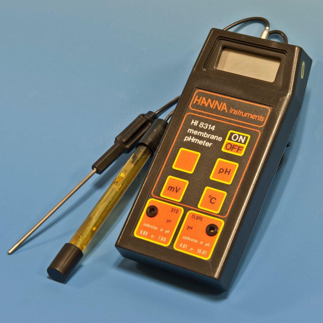 A pH meter and its electrode. Low-pH viral inactivation works by holding the captured antibody pool at a precisely measured acidic pH for a set time — controlling and recording that pH is the whole point of the step.
Laboratory pH meter. Image by Hannes Grobe, CC BY 3.0, via Wikimedia Commons.
A pH meter and its electrode. Low-pH viral inactivation works by holding the captured antibody pool at a precisely measured acidic pH for a set time — controlling and recording that pH is the whole point of the step.
Laboratory pH meter. Image by Hannes Grobe, CC BY 3.0, via Wikimedia Commons.
Our monoclonal antibody (mAb) — a Y-shaped protein drug grown by living cells — is mostly pure now. But it was made by living mammalian cells, almost always CHO cells (Chinese hamster ovary cells) — a rodent line, which is why rodent retroviruses are the canonical virus this step is built to kill — and any process that uses living animal cells carries a tiny theoretical risk: a virus could be hiding in the liquid. Maybe it came in with a raw material years ago. Maybe it was lurking in the cell line itself. The honest answer is that on any given batch we usually find nothing at all — but "usually nothing" is not good enough for a medicine you inject into your bloodstream. And the risk is not purely theoretical: documented bioreactor contamination events — Vesivirus 2117 and Minute Virus of Mice among them — have shut down commercial plants, which is precisely why redundant, validated clearance is mandatory rather than optional. So we do not gamble. We build in deliberate, redundant steps that destroy or remove viruses whether or not any are present. Low-pH inactivation is the first of those steps.
Think about milk. Fresh milk can carry germs, so we pasteurize it: we hold it at a controlled condition for a set amount of time, long enough to kill anything harmful, then move on. Low-pH viral inactivation is the same idea — but instead of heat, we use a brief acid bath, held for a proven length of time, that destroys fragile viruses while leaving our antibody intact.
What this chapter covers
We will walk through what "low pH" actually does to a virus, and why it works on some viruses but not others. We will follow the four-step recipe — drop the pH, hold, destroy, neutralize — and see why the Protein A eluate arrives almost ready for it. Then we will get specific: the real numbers (pH, temperature, hold time, log-reduction), the rules that regulators expect under ICH Q5A, the kinetics that let engineers shorten or lengthen a hold safely, and the choice between an old-fashioned hold tank and a flowing coil. Along the way we will name the standards, vendors, and studies that turn a friendly analogy into a validated manufacturing step.
What actually happens
The pH-hold principle and the chemistry of envelope disruption
First, a quick word on pH. pH is just a number, from 0 to 14, that tells you how acidic or basic a liquid is. Low pH means acidic — lemon juice sits around pH 2, vinegar around pH 3. High pH means basic, like soap. Pure water is neutral at pH 7. The scale is logarithmic, so each whole step is a tenfold change in acid strength. Most viruses, and especially the fragile ones, hate a strongly acidic environment.
Here is the lucky part. The Protein A capture step you just read about releases the antibody by switching to a mild acid, so the antibody flows off the column in an already-acidic liquid called the eluate (the liquid that comes off the column). For a typical platform process that eluate lands somewhere around pH 3.8 to 4.0, depending on the resin and the elution buffer. That means we usually do not have to fight the chemistry — the stream arrives close to the target, and only a small adjustment is needed to push it into the proven inactivation window. That convenience is one reason low-pH inactivation became the industry-standard partner to Protein A capture.
The sequence is simple:
- Adjust the pH down. A small, measured amount of acid is added to bring the liquid to a precise low pH — commonly around pH 3.3 to 3.6. The target is chosen to sit safely below the validated pH ceiling (here pH 3.6) while minimizing acid stress on the antibody. Because the eluate is already acidic, this is a gentle nudge, not a plunge, which keeps the antibody under less stress.
- Hold it there. The acidic stream is held for a proven length of time — the hold time — typically about 60 minutes, though a shorter hold can be valid if it has been demonstrated at a higher temperature. The point is not the exact number on a clock; it is that this specific time, at this pH and temperature, was proven in advance to kill the target viruses. The hold clock starts only after the measured pH — and temperature — reach target across the whole vessel, and it is paused or reset on any excursion; only time spent with both parameters in-window counts toward the proven hold.
- Destroy the virus. Many dangerous viruses wear an outer fatty coat called an envelope. At low pH, hydrogen ions flood the solution and protonate the envelope and capsid (the virus's inner protein shell) proteins, driving them to misfold and unfold irreversibly. Those proteins are the machinery the virus uses to bind and fuse with a host cell, so once they denature — lose that folded shape for good — the virus can no longer infect anything. The lipid envelope is mostly a marker that these viruses are acid-labile — easily broken down by acid: it makes them vulnerable, but the kill is protein denaturation, not lipid lysis. (That is different from solvent/detergent inactivation, where a detergent literally dissolves the lipid coat.) For all practical purposes, the virus is dead.
- Neutralize. A base is added to bring the pH back up to a gentle level. Our antibody, which tolerates the brief acid bath, is now both pure and demonstrably safer, and ready for the next purification step.

The diagram below traces those four steps end to end — eluate in, acid down, hold, neutralize out. The acidic eluate this step inherits is itself a hand-off: the capture step released the antibody with a mild acid, so the stream arrives already part-way into the kill window. Steps 1 and 4 — the acid dose and the base dose — are usually driven by an automated control loop that titrates to a pH setpoint and then holds it, exactly the kind of closed-loop dosing the data trilogy traces in how automation and control data is born.
The science of the kill
Enveloped versus non-enveloped: the orthogonal strategy
Not all viruses are equal in the face of acid, and this is the single most important idea in the chapter. Viruses come in two broad kinds. Enveloped viruses wrap themselves in a stolen scrap of fatty membrane — influenza, herpes, measles, and the retroviruses that can sometimes appear in rodent-derived cell lines all belong to this group. That fatty coat is their weakness: acid tears it apart, so low-pH inactivation is extremely effective against them.
Non-enveloped viruses have no such coat — just a tough protein shell around their genes. The parvoviruses and small enteroviruses fall here, and they shrug off acid almost entirely. To validate a process, engineers deliberately challenge it with a panel of model viruses that spans both classes: the small, hardy non-enveloped Minute Virus of Mice (MVM) and Porcine Parvovirus at the acid-resistant end, alongside enveloped models such as Bovine Viral Diarrhea Virus (BVDV) and the murine (rodent) retroviruses at the acid-susceptible end. Picking viruses from both ends proves the process clears the whole spectrum, not just the easy cases.
This is exactly why a single safety step is never accepted. Low-pH inactivation handles the enveloped viruses brilliantly and the non-enveloped ones barely at all. So a second, completely different method has to cover the gap — and that is the viral filter two chapters from now, which strains viruses out by size and catches the tiny non-enveloped ones acid cannot touch.
| Enveloped | Non-enveloped | |
|---|---|---|
| Coat | A stolen scrap of fatty membrane | Just a tough protein shell |
| Examples | Influenza, herpes, measles, retroviruses, BVDV | Parvoviruses (MVM, PPV), small enteroviruses |
| Acid (low pH) | Susceptible — acid tears the coat apart | Resistant — shrugs acid off |
| Cleared by | Low-pH inactivation (this step) | Viral filtration, by size (a later step) |
Regulators call this pairing orthogonal, and they require it: the international ICH Q5A guideline sets the expectation worldwide [1], and the U.S. FDA adopted that same guidance in January 2024, making the multi-step, orthogonal strategy the regulatory backbone for viral safety under FDA oversight [2].
The real numbers and Arrhenius kinetics
It would be easy to read "pH 3.5 for an hour" as a rule of thumb. It is not. It is a validated specification, anchored in consensus data and dedicated studies.
The number is not a fixed clock — it is whatever a validation study proved at the worst-case corner. Thirty minutes can be plenty at a higher temperature; sixty can be too short if the hold runs cold. The hold time, the pH, and the temperature only mean anything together.
The most concrete reference point is a published standard: ASTM E2888, "Standard Practice for Process for Inactivation of Rodent Retrovirus by pH." It defines a generic, well-characterized operating window: hold at pH 3.6 or below, at 15 °C or above, for more than 30 minutes, and you can expect at least a 4 log₁₀ reduction in rodent retrovirus — meaning the virus count drops by a factor of 10,000 or more [3]. That "log reduction" language is how the whole field talks about kill: 1 log is a 10× drop, 4 logs is 10,000×, and validation studies tally up the logs from every step to prove the total clearance is overwhelmingly larger than any conceivable starting load. Work the arithmetic once and it sticks: if a step is spiked with 10⁷ virus particles and delivers 4 logs, then 10⁷ ÷ 10⁴ = 10³ particles survive. The viral filter's own logs then stack on top. Each step divides the survivors by another factor, and dividing twice is the same as adding the logs — so the total clearance is the sum of each step's logs, not the product of the survivors:
| After | Logs so far (add) | Survivors of a 10⁷ spike |
|---|---|---|
| Spike (start) | 0 | 10,000,000 |
| Low-pH step (4 logs) | 4 | 1,000 |
| Viral filter (4 logs) | 4 + 4 = 8 | under 1 |
Stacking a 4-log step on a 4-log step removes 10,000 × 10,000 = a hundred-million-fold, which in log terms is simply 4 + 4 = 8 logs. That is why you add the logs rather than re-multiplying the surviving particle counts.
Behind that standard sits the science. A landmark 2003 study by Brorson and colleagues at the FDA quantified low-pH inactivation kinetics across many antibody processes and showed that a defined "bracket" of pH, temperature, and time gives reproducible retrovirus clearance of greater than 4.6 logs — strong evidence that the step is a generic, dependable kill rather than something that must be reinvented for every molecule [4]. A later multi-company collaboration pooled real manufacturing data from across the industry and confirmed the same thing: low-pH hold delivers robust, consistent clearance of enveloped viruses across very different processes, and it pairs cleanly with viral filtration as the orthogonal partner [5].
Why does temperature matter so much? Because inactivation is a chemical reaction, and chemical reactions speed up with heat in a way described by Arrhenius kinetics — the warmer the solution, the faster the kill. As a rough rule, every 10 °C rise roughly halves the time needed to reach the same result. That is the lever behind a valid 30-minute hold at a higher temperature versus a 60-minute hold at a cooler one. It is also why temperature is monitored as carefully as pH: a hold run a few degrees colder than validated is a hold that may not have finished its job. To absorb this uncertainty, companies build in a safety factor, validating the step at the worst plausible corner of the window (highest acceptable pH, lowest temperature, shortest time) so that real production, which runs comfortably inside that corner, always over-delivers. Because the kill is a well-behaved function of pH, temperature, and time, those same variables make this step a natural target for a predictive model — a soft sensor (a model that infers a hard-to-measure quantity, here the expected log reduction, from the probe readings you already have): the machine-learning book's viral-safety chapter takes the Arrhenius kinetics and worst-case bracket of this page and turns them into a model that predicts clearance and flags a marginal hold before it fails.
Protecting the antibody: buffer chemistry and aggregate control
There is a tension hiding in all of this. The same acid that destroys a virus can also harm the antibody if we are careless. Held too long, too cold-and-acidic, or in the wrong buffer, a fraction of antibody molecules can partially unfold and clump together into aggregates — clusters that lower yield and, if they reached a patient, could provoke an unwanted immune reaction [6]. So the buffer chemistry is chosen deliberately. Acids like sodium citrate (around pH 3.2) or sodium phosphate (around pH 3.5) are common because they hold the pH steady and minimize how much the antibody is stressed — they must drop the pH into the kill window without stripping the antibody's Fc stem (the stable base of the Y-shaped molecule) or nudging molecules toward hydrophobic — water-repelling — aggregation during the hold. The art of this step is finding the spot that is harsh enough for the virus and mild enough for the drug. The practical levers are concrete: aggregation rises with lower pH, longer hold, and higher protein concentration, and is modulated by the buffer species — so an engineer minimizes time within the validated window, controls the protein concentration, and chooses among the available acids (citrate versus another option such as acetate, for example) to limit clumping [6].
There is a volume cost to watch, too: the acid titrant and any neutralization base add liquid and shift the conductivity of the stream (conductivity is a quick measure of how much dissolved salt the liquid carries; the next polishing column binds the antibody by charge and only loads correctly if the salt level arrives within a set range), so the titrant volume and final conductivity are controlled to keep the pool in spec for the column that follows. Whatever aggregate this hold does create is not the last word: it becomes a defined load that the next chapter's polishing chromatography is designed to scrub back out, which is one reason a small, controlled amount of aggregate formation here is tolerable rather than catastrophic.
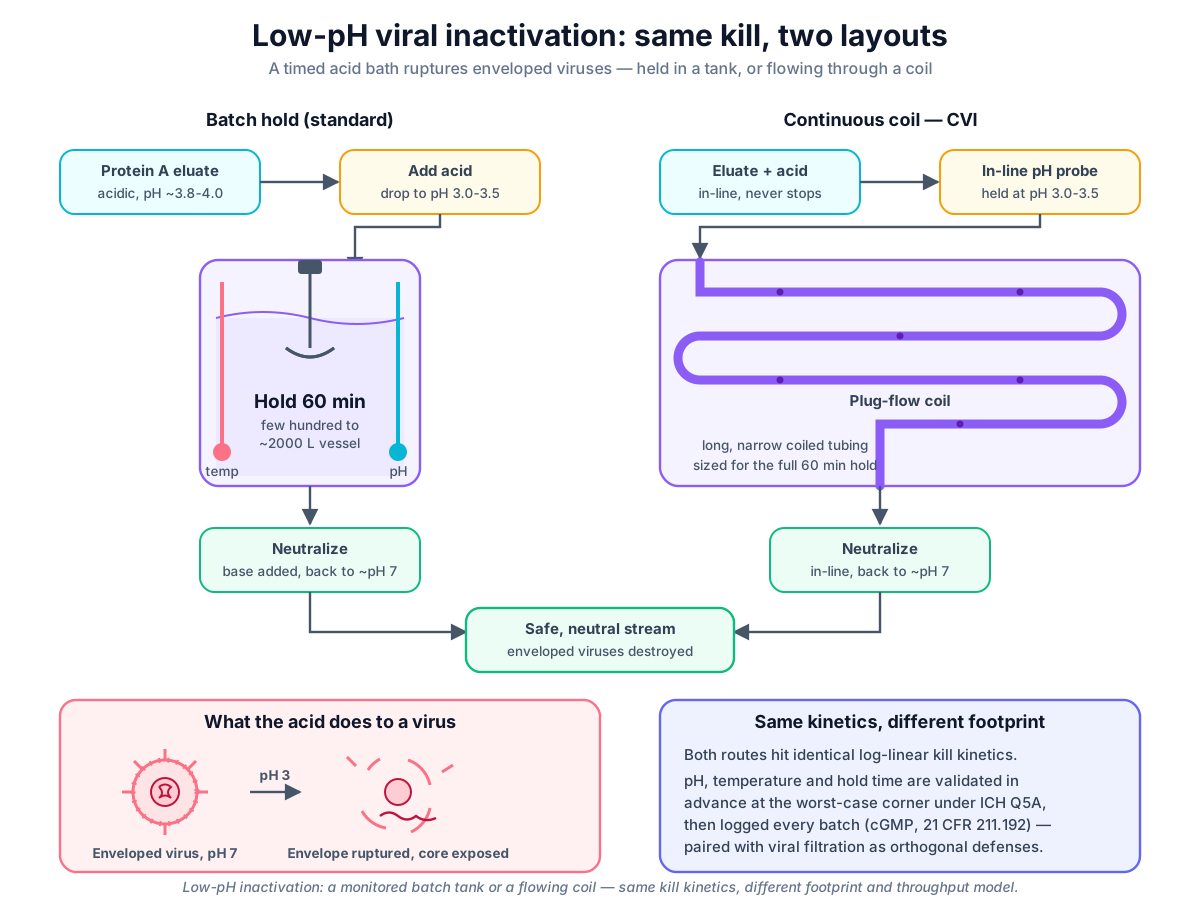 Low-pH inactivation can be performed as batch hold in a monitored tank or continuous residence in a coil; both achieve same kill kinetics but differ in footprint and throughput model
Original diagram by the authors, created with AI assistance.
Low-pH inactivation can be performed as batch hold in a monitored tank or continuous residence in a coil; both achieve same kill kinetics but differ in footprint and throughput model
Original diagram by the authors, created with AI assistance.
Equipment and proof
So far: acid kills enveloped viruses, temperature sets the speed, buffer choice protects the antibody, and everything is proven at the worst-case corner. Now the question turns physical — where does the hold actually happen?
Tank versus coil: equipment design and trade-offs
In the standard commercial process, low-pH inactivation happens in a hold tank: fill a vessel — often a few hundred to a couple thousand liters — add acid while a gentle agitator keeps the contents uniform, watch the pH and temperature for the set time, then neutralize and move on. It is simple, well understood, and used by large manufacturers worldwide. Its weakness is that it is a pause: the whole batch stops and waits while the clock runs.
The modern, continuous approach never stops the flow. Instead of a tank, the acidic stream travels through a long, narrow coil sized so that every drop spends at least the validated hold time inside before it emerges. This is Continuous Viral Inactivation (CVI), and the engineering challenge is subtle: in a simple pipe, liquid in the middle races ahead while liquid at the walls lags, so some drops would exit too soon. Engineers solve this by shaping the flow — a coiled-flow-inverter design repeatedly twists the tube so the liquid mixes across the pipe and every drop's residence time bunches tightly around the target. The foundational engineering of this approach was worked out by Klutz and colleagues at Bayer in 2016 [7], and a later study demonstrated a continuous tubular reactor holding the stream for more than 60 minutes of minimum residence time and matching the retrovirus clearance of a conventional batch hold [8]. It is worth being precise about the history: continuous hold systems and the components behind them have existed in the industry for years; the contribution of newer pilot efforts is integrating them into a fully connected, always-moving line, not inventing the idea.
 Tank and coil reach the same validated kill — they differ in footprint, throughput model, and how the hold time is proven and monitored.
Original diagram by the authors, created with AI assistance.
Tank and coil reach the same validated kill — they differ in footprint, throughput model, and how the hold time is proven and monitored.
Original diagram by the authors, created with AI assistance.
The two layouts also describe themselves differently in equipment terms. A batch tank is a single piece of equipment running a short sequence of steps — fill, hold, neutralize — so its place in a structured equipment-and-batch model is a clean, self-contained unit procedure — the modeling term for one piece of equipment carrying out a bounded sequence of operations (here the fill, hold, and neutralize phases) from start to finish. A continuous coil is a flowing module whose "hold" is a residence-time property of the tubing rather than a timer on a vessel. That distinction matters once the record has to be stored and queried in a structured way, which is exactly the modeling problem the open-source companion works through in its batch and equipment model and its reference architecture. The same structure can be written down as typed relationships — this hold runs-on the hold tank, its pool is derived-from the Protein A eluate, and the inactivation step is-a viral-clearance operation — which is how the ontology book's chapter on relations and genealogy turns a batch into a queryable lineage rather than a pile of free text.
Validation and evidence: how the hold is proven
None of the numbers in this chapter are decided on the plant floor. They are fixed in advance by a dedicated viral-clearance validation study, almost always run at small scale in a specialist biosafety lab rather than in the licensed production area (the GMP manufacturing suite where real batches are made under formal quality rules), because deliberately adding live virus to a production stream is unthinkable. The study spikes a known, high titer (a high concentration of virus) of each model virus into a scaled-down version of the hold, runs the step at the worst-case corner of the window — the highest pH, the lowest temperature, and the shortest time the process will ever be allowed — and then measures how many logs of virus survive. The gap between the spiked load and the survivors is the log reduction value (LRV) claimed for the step, and because it was earned at the worst corner, routine production running comfortably inside that corner always does better. That worst-case-corner logic is why a real batch like the one in the record below holds at pH 3.47 and 18 °C for 62 minutes when this process's validated limits are pH 3.6 or below, 15 °C or above, and 60 minutes or more. ASTM E2888 establishes only a generic floor of more than 30 minutes; this particular running-example process chose to validate a more conservative 60-minute minimum hold, so its envelope is tighter than the standard's. Either way, every routine number sits inside the proven envelope with room to spare.
It is also worth knowing what limits an LRV claim in the first place. The logs you can demonstrate are capped by how much virus you can spike and by the limit of detection of the assay — the laboratory test that counts surviving virus: if the hold drives the count below what the assay can see, the result is reported as "greater than X logs" rather than an exact number. Buffer cytotoxicity and assay interference can further constrain the spike level and recovery, which is why careful study design — high spike titer, large-volume plating — is part of earning a credible claim.
Why it matters
This step does not just clean the product — it protects the patient. A biologic medicine is often injected straight into the body, bypassing every natural barrier, so it must carry no live virus at all. There is no margin for "probably fine."
Regulators — the government agencies that approve medicines — require a documented viral clearance strategy: proof, built from validation studies, that the process can destroy or remove viruses even in a worst case. And they insist it be orthogonal, combining methods that work in completely different ways so each catches what the other misses [1]. Low-pH inactivation is the chemical method, superb against enveloped viruses; the viral filter is the physical method, straining out the rest by size. Two different traps, two different weaknesses covered.
Viral safety is not a place to cut corners. The hold time, pH, and temperature are proven ahead of time in dedicated validation studies, then watched closely on every batch. If the pH is a little too high, the temperature too low, or the hold too short, the step can fail silently — the liquid looks identical, but a virus could survive. That is why these parameters are treated as critical and recorded for every single batch.
In the real world: cGMP records and MES logging
Low-pH inactivation is one of the most universal unit operations in all of antibody manufacturing — nearly every commercial mAb process built on a Protein A platform includes it, precisely because the capture step hands over an acidic stream and the chemistry is so dependable.
The rules around it are global and explicit. The governing document is ICH Q5A, the international guideline on viral safety evaluation of biotechnology products, whose latest revision (R2) reached Step 4 (the ICH stage where a guideline is finalised and recommended for regulators to adopt) in November 2023; it sets the expectation for a multi-step, orthogonal clearance strategy and its validation [1]. As noted earlier, the U.S. FDA adopted that guidance in January 2024, making inactivation a validated, mandated critical step under FDA oversight [2]. And the whole operation runs under cGMP — current Good Manufacturing Practice, the binding U.S. quality rules for making medicines. cGMP is why every batch's pH, temperature, and hold-time trace is captured and reviewed: under 21 CFR 211.192, the production record for each batch must be examined to confirm the step ran inside its validated limits before the batch can proceed [9]. In practice, pH and temperature probes feed a Manufacturing Execution System (MES) that logs readings every few minutes through the hold, and any drift, miscalibration, or excursion becomes a tracked, investigated event rather than a footnote. Because that MES is a computer keeping GMP records, it is itself subject to computer system validation (CSV) — documented proof that the software records and stores the data correctly — increasingly framed as the FDA's leaner, risk-based computer software assurance (CSA), which focuses test effort on the high-risk functions like the hold-time calculation and the quality flag. The electronic record it produces must also meet 21 CFR Part 11 (and its EU equivalent, EudraLex Annex 11): the timestamped pH and temperature trace needs a secure audit trail, attributable user identities, and tamper-evident storage so a reviewer can trust it years later. (Those electronic-record rules are the subject of their own chapter in the data book; this page only notes where they attach.)
Anatomy of a low-pH hold-time record
When the hold finishes, what the batch record actually keeps is not the word "60 minutes." It is a small dossier — an identity card for that one hold — and every field on it is part of the proof that the step did its job. The contemporaneous start timestamp pins the hold to a moment in time. The pH carries both a setpoint (SP) — the value the control loop aimed for — and the process value (PV) actually measured, because the proof lives in the measured trace, not the target. Temperature carries the same SP-versus-PV pair, and its minimum matters most, since a cold dip is what threatens the kill. The hold duration is recorded as the time the stream actually spent with pH and temperature both inside the window, the log-reduction target names the clearance the hold is validated to deliver, and a single quality flag (Good, Uncertain, or Bad) — assigned automatically by the measuring system based on the probe's own health and calibration status — rolls the whole thing up into a verdict that travels with the value. Any moment the stream left the window is logged as a deviation event that opens an investigation.
 Every low-pH hold leaves an identity card, not a lone number: timestamp, pH and temperature setpoint versus measured value, duration, log-reduction target, a quality flag, and any deviation — the contemporaneous proof a reviewer signs off.
Original diagram by the authors, created with AI assistance.
Every low-pH hold leaves an identity card, not a lone number: timestamp, pH and temperature setpoint versus measured value, duration, log-reduction target, a quality flag, and any deviation — the contemporaneous proof a reviewer signs off.
Original diagram by the authors, created with AI assistance.
That bundle — value plus quality flag plus contemporaneous timestamp, never a bare number — is precisely what makes the record trustworthy, and it is the physical seed of a much larger story. In the data trilogy, the same hold-time record is the worked example of ALCOA+ data integrity — an acronym for the qualities a trustworthy record must have: attributable to the operator and QA reviewer, legible, contemporaneous, original, and accurate (the "+" adds completeness, consistency, and enduring availability). The open-source companion then shows the same fields landing as concrete columns and rows that a query can audit.
When the hold fails: real-world deviations and containment
The warning box above is not abstract. The most common ways a low-pH hold fails are quiet, mechanical, and well documented in FDA inspection findings — Form 483 observations (an inspector's list of issues noted at the end of a site inspection) and Warning Letters (the FDA's more serious formal citation of significant violations) that repeatedly fault firms for unreliable monitoring of critical viral-inactivation parameters and for failing to investigate the resulting excursions [10]. Three failure modes recur:
- pH probe miscalibration. If a probe drifts so the displayed pH reads 3.5 while the liquid is really at 3.8, the operator believes the stream is well inside the window when it is sitting above the validated pH 3.6 ceiling. The enveloped-virus kill slows sharply, the batch record looks perfect, and nothing flags — which is exactly why the measured PV, traceable to a calibrated probe, is treated as the evidence rather than the setpoint.
- Temperature-logger drift below 15 °C. Because the kill follows Arrhenius kinetics, a hold a few degrees colder than validated may not finish. A temperature logger that reads high — showing 16 °C while the jacket is really at 13 °C — lets an under-temperature hold pass review. The validated 15 °C floor only protects the batch if the instrument reporting it is trustworthy.
- Coil residence-time tails. In a continuous coil, the failure is geometric rather than electrical: if the flow is not shaped tightly enough, the fastest-moving drops in the center exit before they have spent the full hold time, leaving a tail of under-treated product that blends invisibly into product that was held correctly. This is the specific risk the coiled-flow-inverter design exists to suppress, and why a continuous step is validated on its minimum residence time, not its average.
In every case the antibody solution looks identical afterward — clear, on-spec, indistinguishable — while a virus may have survived. That is what makes the contemporaneous record and its quality flag non-negotiable: the record, reviewed under 21 CFR 211.192 [9], is the only thing standing between a silent excursion and a batch reaching a patient. A logged out-of-window dip becomes a tracked deviation; an unlogged one becomes a data-integrity failure on top of a safety failure.
Key terms
- pH — a number from 0 to 14 describing how acidic (low) or basic (high) a liquid is; each step is a tenfold change in acid strength.
- Eluate — the liquid carrying the antibody after it is washed off a chromatography column; here it arrives already acidic, around pH 3.8 to 4.0.
- Hold time — the proven length of time the stream stays at low pH (typically about 60 minutes, shorter if validated at higher temperature).
- Envelope — the outer fatty coat on some viruses; acid disrupts it, killing the virus.
- Enveloped vs. non-enveloped virus — enveloped viruses (influenza, herpes, retroviruses) have a fatty coat acid can destroy; non-enveloped viruses (parvoviruses, enteroviruses) have only a tough protein shell and resist acid, so they must be removed by filtration.
- Log₁₀ reduction — the unit of viral kill; 1 log is a 10× drop, 5 logs a 100,000× drop.
- Viral clearance — the documented, validated strategy proving the process destroys or removes viruses.
- Orthogonal — using two methods that work in different ways so each catches what the other might miss.
- Arrhenius kinetics — the rule that the inactivation reaction speeds up with temperature; roughly, every 10 °C halves the time needed.
- Aggregate — clumps of partially unfolded antibody that low-pH stress can create; minimized by careful buffer choice.
- Continuous Viral Inactivation (CVI) — doing this step in a flowing coil instead of a tank, sized so every drop gets the full hold time.
- cGMP — current Good Manufacturing Practice; the binding quality rules under which the step is documented and reviewed.
- Setpoint (SP) vs. process value (PV) — the value the control loop aims for versus the value actually measured; the proof of a hold lives in the PV trace, not the SP.
- Log reduction value (LRV) — the number of logs of virus the validation study showed the step removes, earned at the worst-case corner of the window.
- Quality flag — the data-quality verdict (Good, Uncertain, or Bad) carried alongside a measured value so it can never be read in isolation.
- Deviation event — a logged, investigated record of any moment the hold left its validated pH, temperature, or time window.
- Soft sensor — a model that infers a hard-to-measure quantity (here, the expected log reduction) from readings you already have, such as pH and temperature.
- CSV / CSA — computer system validation (proof the software records GMP data correctly) and its leaner, risk-based successor, computer software assurance.
- 21 CFR Part 11 / Annex 11 — the U.S. and EU rules for trustworthy electronic records: audit trails, attributable identities, and tamper-evident storage.
Where this leads
With enveloped viruses destroyed and the stream neutralized, the antibody moves on to polishing chromatography — the next chapter — where one or two more columns scrub away the last traces of impurities, including the broken-virus debris and any antibody aggregates this step may have created, before the second, physical viral-safety barrier later strains out whatever acid could not touch.