What is a biologic? (and what is an antibody?)
📍 Where we are: Stop 2 of 21 — before we build anything, let us understand what we are actually making.
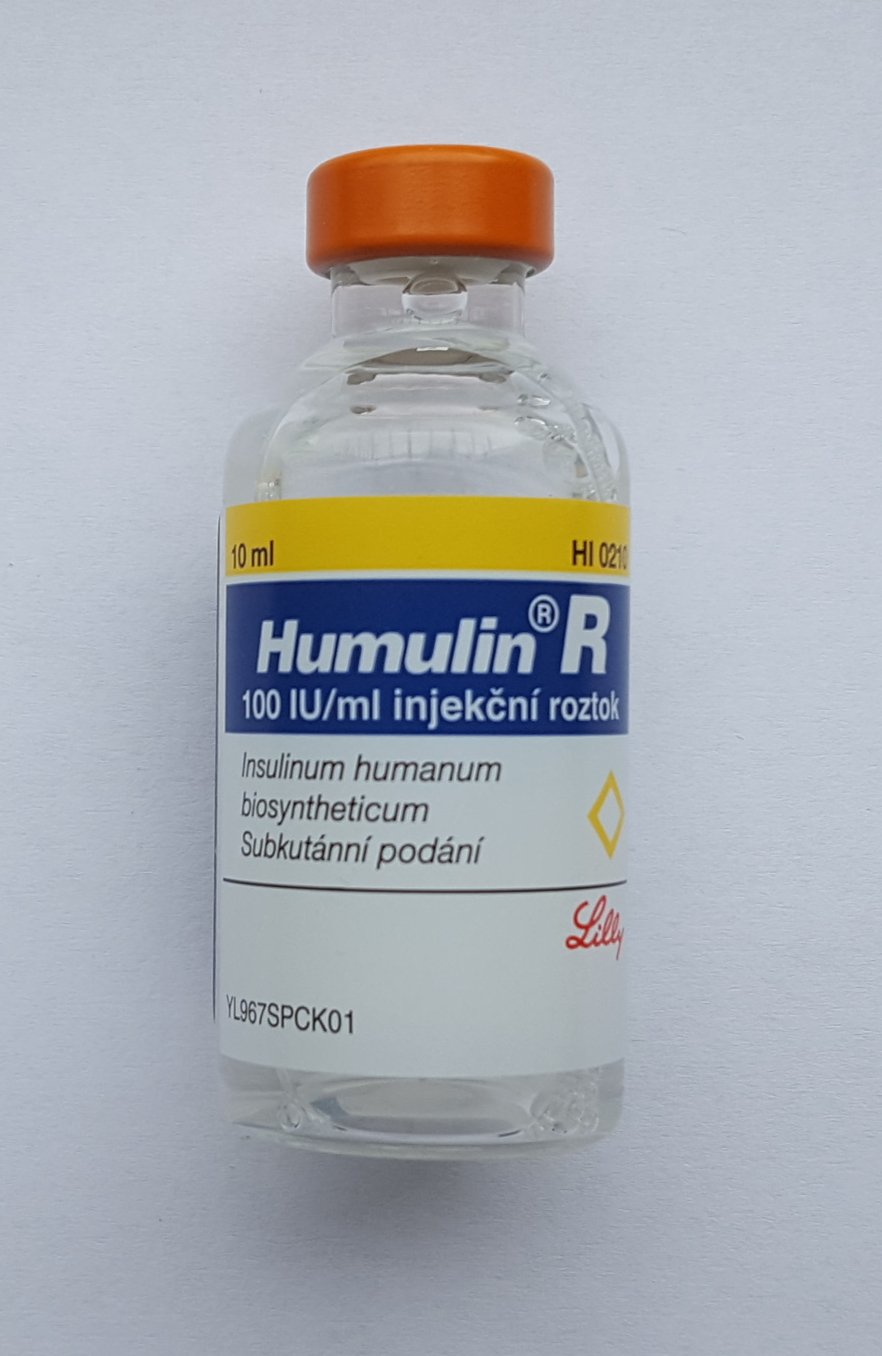 A vial of recombinant human insulin — one of the first biologic medicines. A biologic is a protein drug grown by living cells rather than mixed by chemistry, and this is what one looks like once it reaches the patient.
Human insulin vial. Image by Wesalius, CC BY 4.0, via Wikimedia Commons.
A vial of recombinant human insulin — one of the first biologic medicines. A biologic is a protein drug grown by living cells rather than mixed by chemistry, and this is what one looks like once it reaches the patient.
Human insulin vial. Image by Wesalius, CC BY 4.0, via Wikimedia Commons.
Most pills you know, like aspirin, are tiny chemicals built in a factory. A biologic is different: it is a large, delicate protein (a molecular machine built from chains of building blocks) that can only be made by living cells. This chapter explains what that means, and meets the star of our story: the monoclonal antibody (mAb).
If a normal pill is a bicycle — small, simple, and easy to build the exact same way every time — then an antibody is a jumbo jet made of atoms. Both can carry you somewhere, but one has a handful of parts and the other has hundreds of thousands. You do not build a jumbo jet with the same tools you use for a bike.
What this chapter covers
This chapter zooms all the way in on the medicine itself. First we compare a small-molecule pill with a biologic, so you can feel just how different they are in size and complexity. Then we open up a monoclonal antibody and name its real parts — heavy and light chains, the two gripping arms, the signaling tail, and the tiny sugar decoration that quietly tunes how the body responds. Next we meet the living factory that makes it (the CHO cell), see real numbers for how those cells are grown, and walk through how a single antibody gene becomes a stable production cell line. Along the way we name famous antibodies you have probably heard of, with their makers and approval years, and we ground the whole thing in the rulebooks and the one downstream step — Protein A capture — that nearly every mAb depends on. By the end you will understand the principle that shapes this entire guide: the process is the product.
Small molecule versus biologic
Let us compare the two kinds of medicine, step by step.
A small-molecule drug like aspirin is a tiny chemical, maybe a few dozen atoms. Chemists build it with controlled reactions, like following a baking recipe. Every batch comes out identical, and you can write its exact formula on a napkin. Test the final tablet and you know precisely what you have.
A biologic is enormous by comparison. A typical antibody is a folded protein of roughly 150 kilodaltons (a kilodalton, or kDa, is just a unit of molecular weight) — tens of thousands of atoms, arranged into a precise three-dimensional shape. No chemist can stitch something that large together by hand and have it fold correctly every time. Instead, we hand the genetic instructions to living cells and let them build the protein for us, the way nature builds every protein in your body.
That single fact — grown, not built — has a profound consequence. Because living cells are sensitive, the exact way you grow them becomes part of the medicine. This is captured in the field's founding principle, "the process is the product": for a biologic, the manufacturing process does not merely produce the drug, it largely defines the drug's quality, and so quality must be designed into the process rather than tested in at the end [1].
Anatomy of a monoclonal antibody
Our hero biologic is the monoclonal antibody. The word "antibody" means a Y-shaped protein your immune system makes to recognize one specific target — a virus, a tumor signal, anything it treats as foreign. "Monoclonal" means every single copy is identical, all descended from one original cell, one clone. The idea traces back to the landmark 1975 work of Köhler and Milstein, who fused an antibody-making immune cell (which makes the antibody but dies after a few divisions in culture) with an immortal cell (a tumor cell that divides indefinitely) to create a hybrid line that both keeps making the one antibody and keeps growing forever — secreting one defined antibody, over and over [2]. That is what makes a drug "monoclonal": not a mixture, but one exact molecule produced in vast, uniform quantity.
Now let us open up the Y and name its real parts, because a publication-grade picture needs more than "two arms and a stem."
Structure and function: Fab, Fc, and the variable binding region
An antibody (specifically the IgG class used for most therapeutics) is held together in the familiar Y, with two distinct ends that do two distinct jobs. The two tips of the Y are the Fab regions ("fragment, antigen-binding" — the antigen is simply the target the antibody recognizes). Each Fab ends in a variable region — a stretch that differs from antibody to antibody and is shaped to lock onto exactly one target and nothing else, like a key cut for a single lock. The stem of the Y is the Fc region ("fragment, crystallizable" — the name simply records that this constant tail was the first piece purified antibodies could be made to crystallize into, back when the fragments were first characterized), the constant tail that the immune system reads as a signal: this thing is tagged, deal with it. The variable region decides what the drug grabs; the Fc decides what happens next once it has grabbed.
Heavy and light chains: why four chains make one antibody
That single Y is not one molecule but four protein chains snapped together: two identical heavy chains (the long backbone that runs from a Fab tip all the way down into the Fc stem) and two identical light chains (the shorter chains that cap the outer side of each arm). Inter-heavy-chain disulphide bonds (strong chemical clasps between sulfur atoms on the two chains) at the flexible hinge clasp the two heavy chains together, while a separate disulphide bond links each light chain to its heavy-chain partner near the top of each Fab arm. Because the two halves are mirror images built from identical chains, the finished antibody has two identical binding arms — binding the same target at two points gives the antibody high avidity (a much stronger effective hold than a single arm), and for some targets the bivalency is required to cluster or cross-link the target, not just to bind it more tightly.
Glycosylation at Asn297: where it is, why it matters
One detail matters enormously for how the medicine behaves. Tucked inside the Fc stem, at a specific spot called Asn297 (asparagine — one of the protein's amino-acid building blocks — at position 297 along the heavy chain) in the heavy chain's CH2 domain (one of the folded sections that make up the Fc stem), sits a small chain of sugars — an N-linked glycan. This glycosylation is not decorative. Its precise structure tunes how strongly the Fc engages the immune system, modulating effector functions (the immune actions the Fc triggers) such as ADCC (antibody-dependent cell-mediated cytotoxicity) and CDC (complement-dependent cytotoxicity) [3]. Different cells add slightly different sugars, which is one major reason "the process is the product": change the cell or its growth conditions and you can change Asn297 — and therefore change how the medicine works.
The glycan is best understood not as one molecule but as an identity card with several fields. There is a shared core (two N-acetylglucosamine, or GlcNAc, sugars plus a biantennary mannose trunk — the glycan branches into a Y of two arms, the "antennae," like a tiny tree, with the sugar mannose forming that two-armed trunk) that every copy is built on.
Hanging off that core are the variable fields the process controls: a core fucose that may be present or absent (removing it — afucosylation — sharply raises ADCC); zero, one, or two galactose sugars on the antennae (the G0/G1/G2 labels, with galactosylation influencing CDC); and a usually low level of sialic acid capping the ends (elevated or variable sialylation can shift pharmacokinetics (how the drug moves through the body over time) and clearance (how fast the body removes it) and dampen inflammatory activity, so it too is tracked on the glycan map — not present-but-irrelevant). The combination — the glycoform — is what the body actually reads, and regulators treat that profile as a critical quality attribute (CQA) worth specifying and testing under ICH Q6B [6]. Laid out as the fields of that card:
| Glycan field | What it is | What it tunes |
|---|---|---|
| Core | two GlcNAc sugars plus a mannose trunk (always present) | the fixed base every copy shares |
| Core fucose | present or absent | removing it (afucosylation) sharply raises ADCC |
| Galactose (G0/G1/G2) | zero, one, or two sugars on the antennae | more galactose influences CDC |
| Sialic acid | usually low, caps the ends | shifts pharmacokinetics and clearance; dampens inflammation |
The figure below dissects that card field by field.
 The Asn297 N-glycan as an identity card: a shared core plus tunable fucose, galactose, and sialic-acid fields. Less core fucose raises ADCC; more galactose raises CDC; the cell line and culture conditions set the profile, which is tracked as a critical quality attribute under ICH Q6B.
Original diagram by the authors, created with AI assistance.
The Asn297 N-glycan as an identity card: a shared core plus tunable fucose, galactose, and sialic-acid fields. Less core fucose raises ADCC; more galactose raises CDC; the cell line and culture conditions set the profile, which is tracked as a critical quality attribute under ICH Q6B.
Original diagram by the authors, created with AI assistance.
This is exactly the kind of multi-field artifact that becomes a row of structured data downstream: when the QC lab runs a glycan map (a laboratory test, or assay, that measures the sugar profile), each of these fields turns into a measured number on a release record — the first hint of how a physical molecule becomes a data point you can store, trend, and audit.
 The anatomy of a monoclonal antibody: a Y-shaped protein ~150 kDa in mass, with two identical antigen-binding arms (Fab regions) and one signaling tail (Fc region). The glycosylation site (green) at Asn297 modulates immune response. Every copy from the same cell line is identical.
Original diagram by the authors, created with AI assistance.
The anatomy of a monoclonal antibody: a Y-shaped protein ~150 kDa in mass, with two identical antigen-binding arms (Fab regions) and one signaling tail (Fc region). The glycosylation site (green) at Asn297 modulates immune response. Every copy from the same cell line is identical.
Original diagram by the authors, created with AI assistance.
Here is the same shape drawn as a simple intuition aid, with the real anatomy labels alongside the plain-language ones:

Famous antibodies you have already met
These molecules are not laboratory curiosities — they are household names that have treated millions of people. A few worth knowing, with their generic name (the INN, or International Nonproprietary Name), maker, target, and U.S. approval year:
| Brand | Generic (INN) | Maker | Target | What it does | U.S. approval |
|---|---|---|---|---|---|
| Humira | adalimumab | AbbVie (orig. Abbott) | TNF-alpha | Calms inflammation (rheumatoid arthritis and more) | December 2002 |
| Keytruda | pembrolizumab | Merck | PD-1 | Takes the brakes off the immune system to fight cancer | 2014 (melanoma) |
| Avastin | bevacizumab | Genentech / Roche | VEGF | Starves tumors of blood supply | 2004 |
| Remicade | infliximab | Janssen | TNF | Calms inflammation | 1998 |
Humira was for years the best-selling drug in the world — annual sales peaked at over $20 billion a year (2022) — and Remicade was one of the earliest blockbuster mAbs. You may also remember the COVID antibody treatments used during the pandemic — those, too, were monoclonal antibodies. Every molecule in this table shares the anatomy you just learned, and every one is grown by living cells — which is where we turn next.
Growing and capturing the antibody
CHO cells as the factory: host choice, viral safety, scale
To manufacture an antibody, the industry overwhelmingly turns to one host: CHO cells (Chinese Hamster Ovary cells), the field's reliable workhorse. The great majority of approved monoclonal antibodies and recombinant protein therapeutics are made in CHO, which is why benchmark surveys of the industry treat it as the default production host [4]. There are alternatives — NS0 and SP2/0 (both mouse myeloma lines), PER.C6, and HEK293 (a human line) — but they are far less common for marketed mAbs. CHO dominates partly because it adds human-like glycans (that Asn297 sugar again), grows well in large tanks, and has decades of regulatory track record behind it.
Because we are making medicine inside living cells, we inherit a risk that pure chemistry never faces: the cells, or the raw materials that feed them, could carry a virus. That is why regulators require a formal viral-safety evaluation of any cell-line-derived biologic — testing the cell banks (the qualified frozen stocks of the production cell line that every batch is grown from), and building purification steps that actively remove or inactivate viruses. The governing standard is ICH Q5A(R2) [5]. Viral safety is demonstrated quantitatively as a log reduction value (LRV) summed across orthogonal steps — steps that remove virus by different physical mechanisms, so a particle that slips through one is very unlikely to survive the next — the low-pH hold, Protein A, dedicated viral filtration, and anion exchange — with regulators expecting a cumulative LRV on the order of ≥12 logs (each "log" is a tenfold cut, so ≥12 logs means reducing any virus more than a trillion-fold — removing all but one particle in a trillion) for a model retrovirus. It is a direct consequence of using cells as factories, and we will return to those dedicated viral-clearance steps later in the guide.
Fed-batch culture: duration, cell density, titer targets
How are these cells actually grown? The standard commercial method is fed-batch culture: you fill one large stainless-steel or single-use bioreactor — for commercial mAbs typically 2,000 to 20,000 litres, the size of a small room — periodically feed the cells concentrated nutrients to keep them productive, and then harvest everything once at the end. Some real numbers to anchor the picture:
- A fed-batch run lasts roughly 10 to 14 days (some processes run as long as ~21 days, depending on the cell line and design).
- Peak viable cell density climbs to about 5 to 20 million cells per milliliter (millions of cells in a volume the size of a raindrop).
- A modern, well-tuned process yields a titer — the concentration of antibody in the harvested broth — of about 2 to 8 grams per liter, with high-performing platforms reaching ~10 g/L.
The modern alternative, perfusion culture, keeps fresh medium flowing in and spent medium flowing out continuously, so the cells never sit in their own waste. That pushes density into the range of about 50 to 100 million cells per milliliter and can drive effective titers to 10 grams per liter and beyond over a long run. The rise of these higher titers, and the standardization of CHO fed-batch as the platform, is exactly the industrialization story that reshaped mAb manufacturing [8]. Fed-batch is still the baseline behind most approved products; perfusion and continuous processing are the emerging direction, which we explore as we go.
That final titer is not just a number you wait for — it can be estimated mid-run by a soft sensor (a model that infers a hard-to-measure quantity from the easy live signals) and the feed schedule that drives it can be tuned in far fewer runs by machine learning; the ML book makes both concrete in process development.
From one gene to one clone: transfection, selection, isolation
Where does a "CHO cell that makes our antibody" come from? It does not exist in nature — it has to be engineered, in a stage called cell-line development.
The steps, in plain terms:
- Transfection. We introduce the DNA instructions for our antibody — the heavy-chain and light-chain expression constructs — into CHO cells. A small fraction of cells take up the DNA and integrate it into their own genome.
- Selection. We apply pressure (a selection marker or drug) so that only cells carrying the antibody genes survive and grow.
- Clonal selection. From the survivors, we isolate individual single cells and grow each one into a colony. Some clones produce far more antibody than others, so we screen many of them to find the highest, most stable producers.
- Monoclonality. Because every cell in the final line descends from one single ancestor cell, every antibody molecule it makes is identical. That single-cell origin is what guarantees the product is truly "monoclonal," and it is a regulatory expectation, not just a nicety.
The clone that wins this contest becomes the seed for the entire commercial supply of the drug. Everything downstream — every batch, for years — traces back to it. That "traces back to" is itself a precise, typed relationship — a derived-from edge linking each lot to its parent and ultimately to one frozen cell bank — and when we later draw the whole record as a graph it becomes the genealogy spine the ontology book builds in relations and genealogy. We unpack the full workflow, including the genetic engineering and clone screening, in building the factory cell; here it is enough to see it as one stage in the larger pipeline below.
Protein A affinity capture: the platform step
Once the cells have made the antibody, it has to be pulled out of a messy soup of cells, debris, and impurities. Two impurity classes drive the entire downstream design: host-cell proteins (HCP), the cell's own proteins, which must be cleared to single-digit ppm (parts per million relative to product — fewer than a handful of host-protein molecules for every million antibody molecules, a deliberately strict bar because residual host proteins can provoke an immune reaction in patients), and aggregates — high-molecular-weight clumps of the antibody itself — which a later polishing step removes. (Protein A capture adds one impurity of its own: leached Protein A that bleeds off the resin and must also be cleared.) Almost every monoclonal antibody on the market is purified using the same first step: Protein A affinity chromatography, the industry-standard "platform" capture used for the great majority of marketed mAbs [7].
The trick is elegant. Protein A is a natural molecule that grabs antibodies — and almost nothing else — by their Fc tail (the stem of the Y). Pack it onto tiny beads (a resin) inside a column, pour the harvested broth through, and the antibodies stick while the cell junk washes straight past. Then release the now-purified antibody by dropping to low pH (around pH 3 to 3.5). That acidic hold does double duty — it is deliberately reused as a viral-inactivation step — but it also risks aggregation of the eluted mAb, which is why it is followed by a rapid neutralization back to around pH 5. Because Protein A works for antibody after antibody, it makes a platform process possible — one well-understood recipe reused across products. The resins and systems come from a handful of specialist vendors (names like Cytiva and Repligen), and in the continuous world the single column is replaced by a multi-column arrangement that runs without stopping. We devote a whole chapter to this later.
Step back and the whole path from a gene to a purified molecule is a short, repeatable chain: engineer the cell once, then run it every batch.
 The platform path from one gene to a purified antibody: transfection and cell-line development build the factory cell once per product; fed-batch culture and Protein A capture then run on every batch. Every commercial batch traces back to the single clone chosen in step 2.
Original diagram by the authors, created with AI assistance.
The platform path from one gene to a purified antibody: transfection and cell-line development build the factory cell once per product; fed-batch culture and Protein A capture then run on every batch. Every commercial batch traces back to the single clone chosen in step 2.
Original diagram by the authors, created with AI assistance.
When the glycan quietly shifts: an accepted batch that still loses potency
Here is the failure mode that makes "the process is the product" feel real. Imagine a batch that passes every routine release test — the protein is the right size, it is clean enough, its concentration is on target, and the standard potency assay reads acceptable. Yet a subtle process change — a small pH drift in the bioreactor, a tweak to the feed schedule, or ordinary clone-to-clone variance between cell banks — has nudged the Asn297 profile: a little more core fucose than before, or a little less galactose. Purity and identity look fine. But because afucosylation drives ADCC, that quiet shift can mean reduced ADCC and a real loss of efficacy in patients, even though no obvious specification was breached. This is precisely why the A-Mab case study singled out N-glycosylation as a critical quality attribute and modeled how process inputs move it [10], and why ICH Q6B insists the specification capture the attributes that actually matter, not just the easy-to-measure ones [6].
The lesson is the thread of this whole guide: you cannot test quality into a biologic at the end, because the most consequential properties are built in upstream by living cells. The defense is to watch the process while it runs — every sensor reading, feed addition, and pH trace becomes a signal that the right molecule is being made. Each of those readings is born at a specific instrument and lands as a row in a database, a journey the data book tours in where process data is born; how those signals are then captured and turned into trustworthy records is the subject of its chapters on where instruments and sensors generate data, and the open-source stack that actually stores and serves them — down to the analytical lab that ultimately measures the glycan — is laid out in the reference architecture and the LIMS and ELN chapter. The downstream consequences for the molecule's measured quality — including the glycan map itself — are covered in measuring quality and keeping the protein stable.
Why it matters
Here is the idea that shapes this entire guide: "the process is the product."
With a small molecule, the recipe defines the drug. Two factories following the same formula make the exact same aspirin. But a biologic is so large and complex that the formula alone is not enough. The way the cells fold the protein, the exact sugars they hang on Asn297, the precise growing conditions — all of it becomes part of the final medicine. Change how you make it, and you may change what it actually is.
That is why we cannot make exact copies of biologics the way we make generic pills. The closest we get is a biosimilar — a medicine that is highly similar but never a perfect duplicate, because no one can clone the original maker's exact process.
For patients, this is everything. A small change in shape — or in that Asn297 sugar — can make the medicine weaker, or trigger the body to attack it as foreign. So every step ahead in this guide — every temperature, every filter, every measurement — exists to make the same safe, effective protein, every single time.
In the real world
Because the process defines the product, manufacturers must prove their process is consistent — not just claim it. That proof is mandated by cGMP, current Good Manufacturing Practice: the body of regulations defining how a medicine must be made, documented, and controlled. In the United States these are codified in FDA 21 CFR Part 211, which requires, among much else, that every batch carry a complete, contemporaneous record of exactly how it was made, so any lot can be reconstructed and audited years later. When that record is electronic — as it almost always is today — a companion rule, 21 CFR Part 11, governs the trustworthiness of the data itself, and the watchword is ALCOA+ (every record must be Attributable, Legible, Contemporaneous, Original, and Accurate, plus complete and enduring); the data book devotes its data integrity chapter to exactly that. The "current" in cGMP matters — the standard rises as technology improves, so yesterday's best practice cannot become tomorrow's excuse.
Two more layers of rules shape what "consistent" even means. ICH Q6B defines the specifications — the tests and acceptance criteria for identity, purity, and potency — that characterize a biologic such as a mAb, turning "is this the right molecule, clean enough, and strong enough?" into measurable numbers [6]. And because biosimilars are legally a thing, regulators had to define exactly how similar is similar enough. In the United States a biosimilar is approved under the 351(k) pathway, distinct from the original biologic's 351(a) Biologics License Application (BLA) (the "351" numbers refer to subsections of the Public Health Service Act, the law governing biologics); both demand far more than a generic pill's paperwork. In Europe, the EMA CHMP guideline on biosimilar monoclonal antibodies requires comparability studies — non-clinical and clinical bridging data — to demonstrate that the copy is "highly similar" with no meaningful clinical differences, precisely because identical manufacturing is impossible [9]. The legal standard is "highly similar," never "identical."
The newer way to run the factory — continuous and intensified processing (intensified meaning more product squeezed from a given volume and footprint — higher cell density and titer in a smaller plant), where cells produce nonstop in a perfusion bioreactor and product is captured on a continuous multi-column train — is being demonstrated at pilot scale by efforts across industry and academia. It represents the emerging direction of the field, not today's norm; fed-batch with Protein A still makes the medicines on pharmacy shelves.
Key terms
- Biologic — a large, complex protein medicine made by living cells, not by chemistry alone.
- Small molecule — a tiny chemical drug (like aspirin) built by controlled chemical reactions.
- Protein — a molecular machine folded from chains of building blocks; the body's all-purpose tool.
- Monoclonal antibody (mAb) — a Y-shaped protein, all copies identical, that locks onto one specific target.
- IgG — the antibody class used for most therapeutic mAbs; about 150 kDa, built from two heavy and two light chains.
- Heavy / light chains — the four protein chains (two of each) that make up an antibody.
- Fab region — the two antigen-binding arm tips, containing the variable region that grips one target.
- Fc region — the constant stem of the Y that signals the immune system; carries the Asn297 sugar.
- Glycosylation (Asn297) — the sugar chain at heavy-chain position Asn297 that tunes immune effector function (ADCC/CDC).
- Glycoform — the specific combination of core, fucose, galactose, and sialic acid on the Asn297 glycan; the profile the immune system actually reads.
- ADCC / CDC — antibody-dependent cell-mediated cytotoxicity and complement-dependent cytotoxicity; two immune effector functions the glycan tunes.
- Critical quality attribute (CQA) — a measurable property (such as the glycan profile) that must stay within limits to assure safety or efficacy.
- CHO cell (Chinese Hamster Ovary cell) — the industry's workhorse cell used to manufacture antibodies.
- Cell-line development — engineering CHO cells with the antibody gene and selecting one high-producing clone.
- Titer — the concentration of antibody in the harvested broth, typically 2–8 g/L in modern fed-batch.
- Fed-batch — the standard method: grow, periodically feed, then harvest all at once after ~10–14 days.
- Perfusion — the continuous method: fresh medium in, spent medium out, for higher density and longer runs.
- Protein A capture — the standard affinity step that grabs antibodies by their Fc tail onto resin beads.
- cGMP — current Good Manufacturing Practice; the regulations (in the U.S., 21 CFR Part 211) defining how a medicine must be made, documented, and controlled.
- ALCOA+ — the data-integrity watchword for cGMP records: Attributable, Legible, Contemporaneous, Original, Accurate, plus complete and enduring (governed for electronic records by 21 CFR Part 11).
- Soft sensor — a model that infers a hard-to-measure quantity (such as titer) from the easy, live process signals, rather than from a direct lab measurement.
- The process is the product — the idea that how you make a biologic helps define what it is.
- Biosimilar — a highly similar (but not identical) version of an existing biologic, requiring comparability studies.
Where this leads
You now know what we are making — a large, delicate, sugar-decorated protein that only living cells can build, and that is "monoclonal" because every copy descends from one clone. You also know the one principle that governs everything: the process is the product. Next, in the big picture: upstream, downstream, fill-finish (the bioprocessing overview), we pull back out and lay the whole factory floor end to end — how the "make it" half (upstream) hands off to the "clean it" half (downstream), and how it all finishes in vials. That map is the scaffold every later chapter hangs on, so let us go build it.